Monday, June 10, 2013

Are you in pain?

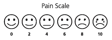
Pain is one of the most difficult things to quantify. The doctor can ask you: How would you classify the pain from 0 to 10, being 10 the most  painful and 0 not painful? and you might answer with a number, but the scale is subjective. So far, there was no method to actually quantify pain. fMRI (functional MRI) might change that.


A recent study found a way to predict the pain intensity with fMRI. Basically, several volunteers (I'm not sure how much did this study paid, but I don't see myself volunteering for a pain study...) were scanned while thermal-induced pain was being applied. Using machine-learning techniques, the scientist found patterns of activity in the brain which increased when pain was applied. In this study, people who are going through break-ups were also invited to participated with the aim of finding common activated regions in physical and "social" pain. Finally, the effect of drugs for pain relieve was also studied.

OK, so now you can quantify my pain, then what? Well, just imagine that your pain prescription is not working so well, because your pain was under or over-estimated... or maybe the source of your pain is not being correctly identified...

There are infinite studies interesting to do, specially ones without any medical interest:
- What hurts more: giving birth or being kicked in the balls?
- Who is suffering more after the break-up: me or my partner? I already can imagine: "Look in this fMRI study how much you made me suffer!"

The full study can be read here:
Wager TD, Atlas LY, Lindquist MA, Roy M, Woo CW, & Kross E (2013). An fMRI-based neurologic signature of physical pain. The New England journal of medicine, 368 (15), 1388-97 PMID: 23574118

Saturday, June 8, 2013

How does a painting look like on X-ray?

The impulse to develop medical imaging comes mainly from two sources: understanding/diagnosing diseases and understanding our body. However, medical imaging technology is sometimes used to study different objects. This post is dedicated to the imaging of one type of these objects: paintings.
  • Why is that? Because with medical imaging we can study the paintings without destroying them. For e.g. it is possible to know which materials were used without taking any sample out.
  • Which technology is used? To study paintings, X-ray technology is mostly used. Infrared technology can also be used, but this is not a typical medical imaging technology. (By the way, MRI is not a good medical technology to study paintings, because the paints have basically no water content.) The most important step to use these technologies was that they become mobile.
  • What else can be seen? Painters have their creative process when painting and it is often possible to understand the previous steps. It is also possible to understand the "mistakes" that the painter made before the final version. These images can even help in the restoration of a painting.
  • What are the differences from imaging a person? Well, the thing here is that dose is not so important, as well as patient movement or "patient impatience" due to long acquisitions.
There are many examples of paintings that were studied with medical imaging. One example of such study was made with the "Sunflowers" from Van Gogh, which can be read here. Mona Lisa has also been scanned and some of Picasso's paintings also, such as "The Old Guitarist" and "Les Demoiselles d'Avignon".

Recognize this painting?

mona lisa emissiograph

More information can be found here and there:
Schreiner, M., Frühmann, B., Jembrih-Simbürger, D., & Linke, R. (2012). X-rays in art and archaeology: An overview Powder Diffraction, 19 (01), 3-11 DOI: 10.1154/1.1649963

Friday, June 7, 2013

Dental Imaging - What is happening there?

The first time I was started thinking about dental imaging from a different point of view was when I heard in a talk the speaker saying: "These are statistics about medical imaging, without dental imaging.". I wondered "Why are dental imaging statistics not included?"

My first thought was "well maybe they are seldom"... then again "why would the speaker referred to them specifically?". Dental imaging is definitely increasing! but it seems these procedures are not accounted for when talking about medical imaging. The main dental imaging procedure is X-ray imaging, because X-rays are great for imaging bones and teeth. The increase comes from the fact that digital imaging is now available, which facilitates handling of images. Also, the widespread of these imaging devices make them more prone to use. When I was young, I made some dental images before I wore braces. These images had to be done in a specific clinical imaging facility and were not performed at the dentist clinic. Now, most dental clinics have these devices. Some even say that you should do an annual check up using this technology, so don't be too surprised if the dentist ask you for this next time.

Should you do it? When is it recommended?
The advantage of doing such type of exam without any complaints, it is probably that you will find a cavity before it becomes painful...or to study the evolution of a problem. The disadvantage are the cost and of course, the dose. Although the small doses present in these exams, they are still there. I refer again to this webtool, where you can calculate on your own the risks: Xrayrisk  Traditionally, dental imaging present intra-oral 2D images, but extra-oral are also possible, including 3D images (tomography) which are obtained with a CT scan. Nevertheless, complex exams such as pan-tomography or tomography should only be done in case there is a problem, not for check up...

The main problem with dental imaging is the lack of regulation (either for prescription or safety) and the training of the human resources for these exams. In imaging clinics, you have human resources with radiography studies and possibly even a physicist. In dental clinical, which human resources do they have? 

For more information about the technology and challenges, this article is a good review:
Vandenberghe B, Jacobs R, & Bosmans H (2010). Modern dental imaging: a review of the current technology and clinical applications in dental practice. European radiology, 20 (11), 2637-55 PMID: 20544352

Tuesday, June 4, 2013

Europe PubMed Central

Yesterday, I have discovered the Europe PubMed Central webpage. This is the European version of the PubMed Central. You can find articles and abstracts, but that you already can find online with Google Scholar. What interested me in this webpage is that you find out more about projects and grants funded in Europe in the tool Grant Lookup. In a time of crisis, not only we want to know how our money is being spent, but also find places where our research might be also important in a time where researchers are the new nomads...

Sunday, June 2, 2013

Procrastination to find the most cited paper in the field of MRI

I have posted recently about the most cited (important?) papers in Medical Imaging in the last ten/five/two years here. Today I look for the most cited papers in the field of MRI. Interesting to note that these 3 papers were published in Neuroimage.

Most cited paper in Radiology, Nuclear Science and Medical Imaging Field about MRI:

- of the last 10 years with 1346 citations:
Ashburner, J., & Friston, K. (2005). Unified segmentation NeuroImage, 26 (3), 839-851 DOI: 10.1016/j.neuroimage.2005.02.018 

This paper is the basis for the SPM framework, one of the most important in the field of MRI. Thus, it is understandable that this paper has a lot of citations, because most researchers who use this framework (and there are a lot, myself included) use this paper in their citations.

of the last 5 years with 250 citations:

Klein, A., Andersson, J., Ardekani, B., Ashburner, J., Avants, B., Chiang, M., Christensen, G., Collins, D., Gee, J., Hellier, P., Song, J., Jenkinson, M., Lepage, C., Rueckert, D., Thompson, P., Vercauteren, T., Woods, R., Mann, J., & Parsey, R. (2009). Evaluation of 14 nonlinear deformation algorithms applied to human brain MRI registration NeuroImage, 46 (3), 786-802 DOI: 10.1016/j.neuroimage.2008.12.037

I have to say that I am quite surprised by finding this paper on top. Brain MRI registration is nowadays considered almost a solved problem and I don't think there are many people looking into this anymore. However, it is always nice to put in your own paper: "I used this registration, because this paper says it is the best".


- of the last 2 years with 106 citations:

Smith, S., Miller, K., Salimi-Khorshidi, G., Webster, M., Beckmann, C., Nichols, T., Ramsey, J., & Woolrich, M. (2011). Network modelling methods for FMRI NeuroImage, 54 (2), 875-891 DOI: 10.1016/j.neuroimage.2010.08.063

I have talked about brain networks some times in this blog and this paper shows me that this topic has been hot in the last two years. This papers discusses different methods to obtain networks with fMRI data: "Many different methods are being used in the literature, but almost none has been carefully validated or compared for use on FMRI timeseries data."

Friday, May 31, 2013

KnowYourImages Youtube Channel

I have posted several videos in this blog and now most of them (27 to be exact) are compiled in a Youtube playlist on the blog's Channel. With this playlist, you don't need to search the blog for the videos, because there are all here. Have fun!


Thursday, May 30, 2013

ESMRMB 2013 - Abstract Submission

This year's deadline for abstract submission is 30th May, so there is no time left to submit. In October, the conference will be in Toulouse, France. Since this is the 30th ESMRMB conference, there will be special surprises according to the organizers. Some preliminary information about the programme is already online.
More information here.


Don't forget that next year, ESMRMB will be held together with ISMRM in Milan and the deadline will be in November. Keep track of your conferences with the Medical Imaging Google Calendar.

Wednesday, May 29, 2013

Angelina Jolie doesn't trust medical imaging

Angelina Jolie shocked us all with her decision to remove both her breasts in order to prevent breast cancer. Her breast cancer risk was calculated based on genetics and was 87%. Now it is 5%. I have to agree that this woman is brave! However, I think a very intense screening could have been done with very good results. There are several methods to detect breast cancer: (digital) mammography, tomosynthesis, breast MRI, ultrasound, positron emission tomography and even microwave imaging. Some without any risk, besides the huge amount of money that they cost if we would perform them on every woman. I think money is not a problem for Angelina Jolie and she could get checked out as many times as she wanted...


The following article "Warner, E. (2004). Surveillance of BRCA1 and BRCA2 Mutation Carriers With Magnetic Resonance Imaging, Ultrasound, Mammography, and Clinical Breast Examination JAMA: The Journal of the American Medical Association, 292 (11), 1317-1325 DOI: 10.1001/jama.292.11.1317" refers all these important points:

- "Women with BRCA1 and BRCA2 mutations who do not undergo prophylactic surgery have a lifetime risk of breast cancer of up to 85%, with a significantly higher risk of breast cancer than the general population from age 25 years onward." - This is the case of Angelina Jolie.

- "The combination of MRI, ultrasound, and mammography had a sensitivity of 95%." - great numbers!

- "To date, the reluctance to use breast MRI for surveillance of high-risk women outside the context of a clinical trial relates, to a large extent, to its high cost and relatively low specificity compared with mammography." - This would not be a problem for Angelina Jolie.

- "In conclusion, our results support the position that MRI-based screening is likely to become the cornerstone of breast cancer surveillance for BRCA1 and BRCA2 mutation carriers, but it is necessary to demonstrate that this surveillance tool lowers breast cancer mortality before it can be recommended for general use." - A public figure like Angelina Jolie could help this matter.

And another article "Kriege M, Brekelmans CT, Boetes C, Besnard PE, Zonderland HM, Obdeijn IM, Manoliu RA, Kok T, Peterse H, Tilanus-Linthorst MM, Muller SH, Meijer S, Oosterwijk JC, Beex LV, Tollenaar RA, de Koning HJ, Rutgers EJ, Klijn JG, & Magnetic Resonance Imaging Screening Study Group (2004). Efficacy of MRI and mammography for breast-cancer screening in women with a familial or genetic predisposition. The New England journal of medicine, 351 (5), 427-37 PMID: 15282350" states: "In conclusion, our study shows that the screening program we used, especially MRI screening, can detect breast cancer at an early stage in women at risk for breast cancer."

Sunday, May 26, 2013

Back online!

After a couple of weeks with important work appointments and then some holidays, I am back online and will start publishing posts as usual.

See you tomorrow!

Friday, May 3, 2013

Microwave for breast imaging?


When we hear the word microwave, we immediately think about the heating device we have in our kitchen. But the word microwave just means waves with wavelengths from ranging from 1 meter to 1 millimeter (corresponding frequencies are 300MHz to 300 GHz). Microwave technology has been used in several engineering fields, and biomedical engineering is no exception. Microwave technology is used in the Radio Frequency components for MRI, but it also can be used as an imaging modality of its own. Microwave Imaging is research in progress, but there have been a number of groups working on this, which makes me believe that this will be available soon.

What are the principles behind Microwave Imaging?
Living tissues have different electrical properties than those of air. Therefore, the setup usually involves a liquid around the object and where the antenna is also immersed, so that the interface is minimized. When microwave radiation interacts with living tissue, microwaves can be transmitted, reflected or dissipated (by heat). The penetration of the microwaves goes until about 10 cm. The transmitting antenna emits microwaves and other antennas receive the transmitted and reflected waves. (The transmitting antenna can also receive.) There are differences in the properties of healthy tissue and malignant tumors. The tumors cause the waves to suffer scatter and the waves have now different energy (frequency). With the information of the detected waves, an image can be obtained (through some complicated methods) or the signals can be analyzed.

Advantages of Microwave Imaging:
- Low Cost
- Non-ionizing radiation


The main clinical application of Microwave imaging is breast cancer. This is due to several reasons:
1. Breast tissues are usually above the 10 cm penetration limit.
2. Breast cancer screening will greatly benefit from a low cost and non-ionizing radiation, as well as less painful acquisition for the patients.


Image from here.

More about this technique can be read in:
Fear, E., Meaney, P., & Stuchly, M. (2003). Microwaves for breast cancer detection? IEEE Potentials, 22 (1), 12-18 DOI: 10.1109/MP.2003.1180933
Nikolova, N. (2011). Microwave Imaging for Breast Cancer IEEE Microwave Magazine, 12 (7), 78-94 DOI: 10.1109/MMM.2011.942702

Recent news about microwave imaging for breast cancer:
http://www.sciencedaily.com/releases/2013/04/130423211830.htm

Thursday, May 2, 2013

Dissecting Art, Intersecting Anatomy - Medical illustration

Pauline Lariviere was an artist whose main contributions were made to the field of medical illustration. As a scientist in the medical field, medical illustrations are essential to education. I have already spent some hours drawing something in the computer for a paper or presentation. In old times, all illustrations were done by hand, but nowadays computers play an important role and medical images are often used as a basis to medical illustrations. Here is an example of a medical illustration based on a CT scan:
figure

The video is about an exhibition which pays a tribute to Pauline Lariviere.

Dissecting Art; Intersecting Anatomy from Phillip Schalekamp on Vimeo.

More about the history of medical illustration and the role of computers can be read in:
Tsafrir, J., & Ohry, A. (2001). Medical illustration: from caves to cyberspace‡ Health Information & Libraries Journal, 18 (2), 99-109 DOI: 10.1046/j.1471-1842.2001.d01-16.x
Corl, F., Garland, M., & Fishman, E. (2000). Role of Computer Technology in Medical Illustration American Journal of Roentgenology, 175 (6), 1519-1524 DOI: 10.2214/ajr.175.6.1751519

Wednesday, May 1, 2013

European Month of the Brain

Today starts the European Month of the Brain. I certainly hoped that this information would have been publicized much more. I only come to this information by chance. Let's hope we hear more about this during this month!

"Brain research in Europe needs a decisive effort. We must invest more, define clearer and more innovative research and healthcare strategies, and better coordinate national efforts. We therefore decided to make May 2013 the European Month of the Brain - a full month packed with events and activities to make decision-makers, stakeholders, the media and the public aware of the successes achieved and the challenges still laying ahead for brain research."

http://ec.europa.eu/research/conferences/2013/brain-month/images/brainMonth_medium-print_tag.jpg

The main webpage is here and links about funding are also very useful.

Tuesday, April 30, 2013

What does the Muse CD cover have to do with Medical Imaging?

This is Muse CD cover of their 6th album: "The 2nd Law":

Indeed, this image is truly beautiful and remarkable. It is an image of the white matter fibers in the brain obtained with diffusion MRI (link). The image was obtained by the Human Connectome Project, which is a 5-year project funded by NIH to find the networks of the human brain. These networks will show how our brain communicates between different regions and give insight about the anatomical and functional organization of the brain. The project also has the goal to produce data that will help understanding brain diseases such as Alzheimer's disease. The data is available to the scientific community.

So how do you obtain these networks? By applying computer algorithms to data obtained with different neuroimaging techniques: MRI, fMRI, diffusion MRI among others. These computer algorithms come from the graph theory. The application of these algorithms is extremely useful, because the algorithms analyze the network, reduce the complexity, find similarities and differences between different networks.

A very nice science article for researchers not familiar with the topic:
http://www.sciencemag.org/site/products/lst_20130118.xhtml

To know more about obtaining diffusion MRI data or network methods, look into these two articles:
- Hasan, K., Walimuni, I., Abid, H., & Hahn, K. (2011). A review of diffusion tensor magnetic resonance imaging computational methods and software tools Computers in Biology and Medicine, 41 (12), 1062-1072 DOI: 10.1016/j.compbiomed.2010.10.008
- Kaiser, M. (2011). A tutorial in connectome analysis: Topological and spatial features of brain networks NeuroImage, 57 (3), 892-907 DOI: 10.1016/j.neuroimage.2011.05.025

Monday, April 29, 2013

CT scans help Anthropology studies

This video has caught my attention few days ago. Field Museum in Chicago has used a CT scan to help them with the face reconstruction of an ancient skull (12,000 to 15,000 year old). The great advantage of using a CT scan is that once you have scanned the skull, you can do the processing and reconstruction of the skull on a computer without having to touch it, which means that the skull is never damaged. The problem here is usually the transport of the skull to the CT scan, which are usually located in hospital or clinics. This time, they used a mobile CT scan, which travels to the museum instead of the skull having to travel. Moreover, the researchers used a 3D printer to obtain a model of the skull, which was then used by a forensic artist to do the final face reconstruction. The motivation behind this new scan was that the researchers were not happy with the face reconstruction done before, which made this skull look "too Neanderthal".

After watching this video, I have look for publications which describe the techniques used, but I haven't found many detailed publications. However, I found this review publication (Lynnerup, N. (2010). Medical Imaging of Mummies and Bog Bodies – A Mini-Review Gerontology, 56 (5), 441-448 DOI: 10.1159/000266031), which I have read and although not too detailed about the CT scanning procedures, I have learned a few things:
- Living tissues require different processing that dead tissues, because Hounsfield units are different. Bone tends to de-mineralize, while soft tissues tend to become denser (probably due to surrounding minerals).
- Even that this type of scans are done since the 70's, new CT scans are also done, because technology has improved that new findings arise.
- CT data can be easily shared with other scientists, while skulls and mummies can't.


Saturday, April 27, 2013

Procrastination to find the most cited papers in Medical Imaging

Few day ago, I was wondering what were the most cited (important?) papers in Medical Imaging in the last ten/five/two years. The problem was that I didn't know exactly how to find this information. I googled a bit around and I found a way and tried it out. I found also some extra information about the subject:

Published Papers in Radiology, Nuclear Science and Medical Imaging Field:

Citations in Radiology, Nuclear Science and Medical Imaging Field:

It is interesting to note that it is increasing steadily every year. 

Most cited paper in Radiology, Nuclear Science and Medical Imaging Field:

- of the last 10 years
Jan, S., Santin, G., Strul, D., Staelens, S., Assié, K., Autret, D., Avner, S., Barbier, R., Bardiès, M., Bloomfield, P., Brasse, D., Breton, V., Bruyndonckx, P., Buvat, I., Chatziioannou, A., Choi, Y., Chung, Y., Comtat, C., Donnarieix, D., Ferrer, L., Glick, S., Groiselle, C., Guez, D., Honore, P., Kerhoas-Cavata, S., Kirov, A., Kohli, V., Koole, M., Krieguer, M., Laan, D., Lamare, F., Largeron, G., Lartizien, C., Lazaro, D., Maas, M., Maigne, L., Mayet, F., Melot, F., Merheb, C., Pennacchio, E., Perez, J., Pietrzyk, U., Rannou, F., Rey, M., Schaart, D., Schmidtlein, C., Simon, L., Song, T., Vieira, J., Visvikis, D., Walle, R., Wieërs, E., & Morel, C. (2004). GATE: a simulation toolkit for PET and SPECT Physics in Medicine and Biology, 49 (19), 4543-4561 DOI: 10.1088/0031-9155/49/19/007

- of the last 5 years
Klein, S., Staring, M., Murphy, K., Viergever, M., & Pluim, J. (2010). elastix: A Toolbox for Intensity-Based Medical Image Registration IEEE Transactions on Medical Imaging, 29 (1), 196-205 DOI: 10.1109/TMI.2009.2035616


- of the last 2 years
Hricak, H., Brenner, D., Adelstein, S., Frush, D., Hall, E., Howell, R., McCollough, C., Mettler, F., Pearce, M., Suleiman, O., Thrall, J., & Wagner, L. (2010). Managing Radiation Use in Medical Imaging: A Multifaceted Challenge Radiology, 258 (3), 889-905 DOI: 10.1148/radiol.10101157

How to find this information?
Go to Web of Knowledge and follow these tutorial:
http://www.umuc.edu/library/libhow/woshighlycited_tutorial.cfm

Friday, April 26, 2013

The best blogs in Neuroscience

With the new community of Research Blogging, I have come across with 4 blogs, which are probably the most famous and known in the neuroscience blogosphere of research blogging:


Image from here.

The Scicurious Brain: the Good, Bad, and  Weird in Psychology and Neuroscience and my recommended articles are:
- Beer, Dopamine and Brain Scans
- The BRAIN initiative: BAM or BUST?

Neuroskeptic and my recommended articles are:
- The Man with Uncrossed Eyes
- What is wrong with "Publish or Perish"?

Neurocritic and my recommended articles are:
How Neuroscientists Scan the Media
- What Is This Thing Called Neuroscience?

Brain Posts and my recommendation articles are:
- DTI identifies Brain Aging Changes
- Blast-related Traumatic Brain Injury

These blogs are from this moment in the blog list on the right side of this blog. Did you already know these blogs? Or know other very good ones?

Thursday, April 25, 2013

Research Blogging

This blog is now part of the community in researchblogging.org. "ResearchBlogging.org is a system for identifying the best, most thoughtful blog posts about peer-reviewed research." Know Your Images is now one of the blogs which ResearchBlogging checks for posts about peer-reviewed articles. Furthermore, it is a great way to find new blogs and also keeps you updated about the blogs you like.

Of course, this blog will keep a fare amount of posts which are not dedicated to peer-reviewed articles, but which are just fun to know about and watch.

Wednesday, April 24, 2013

More art inspired by Medical imaging

Marilene Oliver is another artist who is inspired by medical images. She creates sculptures and installations based on medical images. The full portfolio can be seen here. I show in this post some images of her most interesting work:


Tuesday, April 23, 2013

fMRI lie detection and the Semrau case

Semrau is a psychologist accused of committing fraud to Medicare and Medicaid. The case became mostly famous, because he asked that fMRI lie detection would be a evidence in court. The judge had to decide if fMRI was admissible and after hearing scientists advocating for both sides, he has decided not to admit such evidence. However, the question is: Will it be possible to use fMRI lie detection one day?, because the reason for not admitting it has been based on the error rates and acceptance by scientific community and that can change any day...


http://dericbownds.net/uploaded_images/duped.jpg
Image from here

So how does fMRI lie detection work at the moment?
- A deception task is presented to the volunteers: they have to lie about the object they have taken from a box (or similar, such as a card from envelope).
- The volunteer goes inside the scanner and structural MRI is performed and a motor task can also be performed to make the volunteers more familiar with the MRI itself.
- The deception task starts and the volunteer is asked questions about the stolen object among other questions. The volunteer has to lie about stealing the object. During this time, EPI (Echo Planar Imaging) images are acquired.
- Processing of data starts, which includes reorientation and motion correction. Brain patterns are analyzed to detect lying.

Findings have shown that there are specific activated areas (anterior cingulate and the prefrontal cortex) in subjects in the task of deception when a group study is performed. This is a group study, but for fMRI to become a lie detector, it has to stand in individual studies. This has been difficult, because fMRI is a technique with a low signal-to-noise ratio, but some studies have been done. Moreover, deception tasks in these studies are still simple ones, while more complex ones (like the Semrau case) have not been performed.

One of the studies which presented results on individual basis (the one referenced here at the bottom) has led that a company has been formed to sell this type of service (CEPHOS). This was the company involved in the Semrau case and the CEO of this company is the scientist advocating for the fMRI lie detection. The two scientists which advocated against the fMRI lie detector were Marc Raichle, PhD (Wash. U. St. Louis, Neuroscience) and Peter Imrey, PhD (Cleveland Clinic, Statistics).

Anyway, my personal belief is that fMRI should be on the service of health and not of law...

Other Links:
http://www.sciencemag.org/content/328/5984/1336.1.full
http://news.sciencemag.org/scienceinsider/2010/05/fmri-lie-detection-gets-its-day-.html
http://blogs.law.stanford.edu/lawandbiosciences/2012/09/07/hot-news-6th-circuit-affirms-in-us-v-semrau-says-no-to-fmri-lie-detection/

Kozel, F., Johnson, K., Mu, Q., Grenesko, E., Laken, S., & George, M. (2005). Detecting Deception Using Functional Magnetic Resonance Imaging Biological Psychiatry, 58 (8), 605-613 DOI: 10.1016/j.biopsych.2005.07.040