Monday, June 10, 2013

Are you in pain?

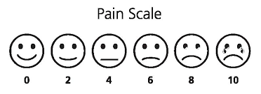
Pain is one of the most difficult things to quantify. The doctor can ask you: How would you classify the pain from 0 to 10, being 10 the most  painful and 0 not painful? and you might answer with a number, but the scale is subjective. So far, there was no method to actually quantify pain. fMRI (functional MRI) might change that.


A recent study found a way to predict the pain intensity with fMRI. Basically, several volunteers (I'm not sure how much did this study paid, but I don't see myself volunteering for a pain study...) were scanned while thermal-induced pain was being applied. Using machine-learning techniques, the scientist found patterns of activity in the brain which increased when pain was applied. In this study, people who are going through break-ups were also invited to participated with the aim of finding common activated regions in physical and "social" pain. Finally, the effect of drugs for pain relieve was also studied.

OK, so now you can quantify my pain, then what? Well, just imagine that your pain prescription is not working so well, because your pain was under or over-estimated... or maybe the source of your pain is not being correctly identified...

There are infinite studies interesting to do, specially ones without any medical interest:
- What hurts more: giving birth or being kicked in the balls?
- Who is suffering more after the break-up: me or my partner? I already can imagine: "Look in this fMRI study how much you made me suffer!"

The full study can be read here:
Wager TD, Atlas LY, Lindquist MA, Roy M, Woo CW, & Kross E (2013). An fMRI-based neurologic signature of physical pain. The New England journal of medicine, 368 (15), 1388-97 PMID: 23574118

Saturday, June 8, 2013

How does a painting look like on X-ray?

The impulse to develop medical imaging comes mainly from two sources: understanding/diagnosing diseases and understanding our body. However, medical imaging technology is sometimes used to study different objects. This post is dedicated to the imaging of one type of these objects: paintings.
  • Why is that? Because with medical imaging we can study the paintings without destroying them. For e.g. it is possible to know which materials were used without taking any sample out.
  • Which technology is used? To study paintings, X-ray technology is mostly used. Infrared technology can also be used, but this is not a typical medical imaging technology. (By the way, MRI is not a good medical technology to study paintings, because the paints have basically no water content.) The most important step to use these technologies was that they become mobile.
  • What else can be seen? Painters have their creative process when painting and it is often possible to understand the previous steps. It is also possible to understand the "mistakes" that the painter made before the final version. These images can even help in the restoration of a painting.
  • What are the differences from imaging a person? Well, the thing here is that dose is not so important, as well as patient movement or "patient impatience" due to long acquisitions.
There are many examples of paintings that were studied with medical imaging. One example of such study was made with the "Sunflowers" from Van Gogh, which can be read here. Mona Lisa has also been scanned and some of Picasso's paintings also, such as "The Old Guitarist" and "Les Demoiselles d'Avignon".

Recognize this painting?

mona lisa emissiograph

More information can be found here and there:
Schreiner, M., Frühmann, B., Jembrih-Simbürger, D., & Linke, R. (2012). X-rays in art and archaeology: An overview Powder Diffraction, 19 (01), 3-11 DOI: 10.1154/1.1649963

Friday, June 7, 2013

Dental Imaging - What is happening there?

The first time I was started thinking about dental imaging from a different point of view was when I heard in a talk the speaker saying: "These are statistics about medical imaging, without dental imaging.". I wondered "Why are dental imaging statistics not included?"

My first thought was "well maybe they are seldom"... then again "why would the speaker referred to them specifically?". Dental imaging is definitely increasing! but it seems these procedures are not accounted for when talking about medical imaging. The main dental imaging procedure is X-ray imaging, because X-rays are great for imaging bones and teeth. The increase comes from the fact that digital imaging is now available, which facilitates handling of images. Also, the widespread of these imaging devices make them more prone to use. When I was young, I made some dental images before I wore braces. These images had to be done in a specific clinical imaging facility and were not performed at the dentist clinic. Now, most dental clinics have these devices. Some even say that you should do an annual check up using this technology, so don't be too surprised if the dentist ask you for this next time.

Should you do it? When is it recommended?
The advantage of doing such type of exam without any complaints, it is probably that you will find a cavity before it becomes painful...or to study the evolution of a problem. The disadvantage are the cost and of course, the dose. Although the small doses present in these exams, they are still there. I refer again to this webtool, where you can calculate on your own the risks: Xrayrisk  Traditionally, dental imaging present intra-oral 2D images, but extra-oral are also possible, including 3D images (tomography) which are obtained with a CT scan. Nevertheless, complex exams such as pan-tomography or tomography should only be done in case there is a problem, not for check up...

The main problem with dental imaging is the lack of regulation (either for prescription or safety) and the training of the human resources for these exams. In imaging clinics, you have human resources with radiography studies and possibly even a physicist. In dental clinical, which human resources do they have? 

For more information about the technology and challenges, this article is a good review:
Vandenberghe B, Jacobs R, & Bosmans H (2010). Modern dental imaging: a review of the current technology and clinical applications in dental practice. European radiology, 20 (11), 2637-55 PMID: 20544352

Tuesday, June 4, 2013

Europe PubMed Central

Yesterday, I have discovered the Europe PubMed Central webpage. This is the European version of the PubMed Central. You can find articles and abstracts, but that you already can find online with Google Scholar. What interested me in this webpage is that you find out more about projects and grants funded in Europe in the tool Grant Lookup. In a time of crisis, not only we want to know how our money is being spent, but also find places where our research might be also important in a time where researchers are the new nomads...

Sunday, June 2, 2013

Procrastination to find the most cited paper in the field of MRI

I have posted recently about the most cited (important?) papers in Medical Imaging in the last ten/five/two years here. Today I look for the most cited papers in the field of MRI. Interesting to note that these 3 papers were published in Neuroimage.

Most cited paper in Radiology, Nuclear Science and Medical Imaging Field about MRI:

- of the last 10 years with 1346 citations:
Ashburner, J., & Friston, K. (2005). Unified segmentation NeuroImage, 26 (3), 839-851 DOI: 10.1016/j.neuroimage.2005.02.018 

This paper is the basis for the SPM framework, one of the most important in the field of MRI. Thus, it is understandable that this paper has a lot of citations, because most researchers who use this framework (and there are a lot, myself included) use this paper in their citations.

of the last 5 years with 250 citations:

Klein, A., Andersson, J., Ardekani, B., Ashburner, J., Avants, B., Chiang, M., Christensen, G., Collins, D., Gee, J., Hellier, P., Song, J., Jenkinson, M., Lepage, C., Rueckert, D., Thompson, P., Vercauteren, T., Woods, R., Mann, J., & Parsey, R. (2009). Evaluation of 14 nonlinear deformation algorithms applied to human brain MRI registration NeuroImage, 46 (3), 786-802 DOI: 10.1016/j.neuroimage.2008.12.037

I have to say that I am quite surprised by finding this paper on top. Brain MRI registration is nowadays considered almost a solved problem and I don't think there are many people looking into this anymore. However, it is always nice to put in your own paper: "I used this registration, because this paper says it is the best".


- of the last 2 years with 106 citations:

Smith, S., Miller, K., Salimi-Khorshidi, G., Webster, M., Beckmann, C., Nichols, T., Ramsey, J., & Woolrich, M. (2011). Network modelling methods for FMRI NeuroImage, 54 (2), 875-891 DOI: 10.1016/j.neuroimage.2010.08.063

I have talked about brain networks some times in this blog and this paper shows me that this topic has been hot in the last two years. This papers discusses different methods to obtain networks with fMRI data: "Many different methods are being used in the literature, but almost none has been carefully validated or compared for use on FMRI timeseries data."