Showing posts with label fMRI. Show all posts

Monday, June 10, 2013

Are you in pain?

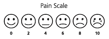
Pain is one of the most difficult things to quantify. The doctor can ask you: How would you classify the pain from 0 to 10, being 10 the most  painful and 0 not painful? and you might answer with a number, but the scale is subjective. So far, there was no method to actually quantify pain. fMRI (functional MRI) might change that.


A recent study found a way to predict the pain intensity with fMRI. Basically, several volunteers (I'm not sure how much did this study paid, but I don't see myself volunteering for a pain study...) were scanned while thermal-induced pain was being applied. Using machine-learning techniques, the scientist found patterns of activity in the brain which increased when pain was applied. In this study, people who are going through break-ups were also invited to participated with the aim of finding common activated regions in physical and "social" pain. Finally, the effect of drugs for pain relieve was also studied.

OK, so now you can quantify my pain, then what? Well, just imagine that your pain prescription is not working so well, because your pain was under or over-estimated... or maybe the source of your pain is not being correctly identified...

There are infinite studies interesting to do, specially ones without any medical interest:
- What hurts more: giving birth or being kicked in the balls?
- Who is suffering more after the break-up: me or my partner? I already can imagine: "Look in this fMRI study how much you made me suffer!"

The full study can be read here:
Wager TD, Atlas LY, Lindquist MA, Roy M, Woo CW, & Kross E (2013). An fMRI-based neurologic signature of physical pain. The New England journal of medicine, 368 (15), 1388-97 PMID: 23574118

Sunday, June 2, 2013

Procrastination to find the most cited paper in the field of MRI

I have posted recently about the most cited (important?) papers in Medical Imaging in the last ten/five/two years here. Today I look for the most cited papers in the field of MRI. Interesting to note that these 3 papers were published in Neuroimage.

Most cited paper in Radiology, Nuclear Science and Medical Imaging Field about MRI:

- of the last 10 years with 1346 citations:
Ashburner, J., & Friston, K. (2005). Unified segmentation NeuroImage, 26 (3), 839-851 DOI: 10.1016/j.neuroimage.2005.02.018 

This paper is the basis for the SPM framework, one of the most important in the field of MRI. Thus, it is understandable that this paper has a lot of citations, because most researchers who use this framework (and there are a lot, myself included) use this paper in their citations.

of the last 5 years with 250 citations:

Klein, A., Andersson, J., Ardekani, B., Ashburner, J., Avants, B., Chiang, M., Christensen, G., Collins, D., Gee, J., Hellier, P., Song, J., Jenkinson, M., Lepage, C., Rueckert, D., Thompson, P., Vercauteren, T., Woods, R., Mann, J., & Parsey, R. (2009). Evaluation of 14 nonlinear deformation algorithms applied to human brain MRI registration NeuroImage, 46 (3), 786-802 DOI: 10.1016/j.neuroimage.2008.12.037

I have to say that I am quite surprised by finding this paper on top. Brain MRI registration is nowadays considered almost a solved problem and I don't think there are many people looking into this anymore. However, it is always nice to put in your own paper: "I used this registration, because this paper says it is the best".


- of the last 2 years with 106 citations:

Smith, S., Miller, K., Salimi-Khorshidi, G., Webster, M., Beckmann, C., Nichols, T., Ramsey, J., & Woolrich, M. (2011). Network modelling methods for FMRI NeuroImage, 54 (2), 875-891 DOI: 10.1016/j.neuroimage.2010.08.063

I have talked about brain networks some times in this blog and this paper shows me that this topic has been hot in the last two years. This papers discusses different methods to obtain networks with fMRI data: "Many different methods are being used in the literature, but almost none has been carefully validated or compared for use on FMRI timeseries data."

Tuesday, April 30, 2013

What does the Muse CD cover have to do with Medical Imaging?

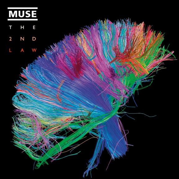
This is Muse CD cover of their 6th album: "The 2nd Law":

Indeed, this image is truly beautiful and remarkable. It is an image of the white matter fibers in the brain obtained with diffusion MRI (link). The image was obtained by the Human Connectome Project, which is a 5-year project funded by NIH to find the networks of the human brain. These networks will show how our brain communicates between different regions and give insight about the anatomical and functional organization of the brain. The project also has the goal to produce data that will help understanding brain diseases such as Alzheimer's disease. The data is available to the scientific community.

So how do you obtain these networks? By applying computer algorithms to data obtained with different neuroimaging techniques: MRI, fMRI, diffusion MRI among others. These computer algorithms come from the graph theory. The application of these algorithms is extremely useful, because the algorithms analyze the network, reduce the complexity, find similarities and differences between different networks.

A very nice science article for researchers not familiar with the topic:
http://www.sciencemag.org/site/products/lst_20130118.xhtml

To know more about obtaining diffusion MRI data or network methods, look into these two articles:
- Hasan, K., Walimuni, I., Abid, H., & Hahn, K. (2011). A review of diffusion tensor magnetic resonance imaging computational methods and software tools Computers in Biology and Medicine, 41 (12), 1062-1072 DOI: 10.1016/j.compbiomed.2010.10.008
- Kaiser, M. (2011). A tutorial in connectome analysis: Topological and spatial features of brain networks NeuroImage, 57 (3), 892-907 DOI: 10.1016/j.neuroimage.2011.05.025

Tuesday, April 23, 2013

fMRI lie detection and the Semrau case

Semrau is a psychologist accused of committing fraud to Medicare and Medicaid. The case became mostly famous, because he asked that fMRI lie detection would be a evidence in court. The judge had to decide if fMRI was admissible and after hearing scientists advocating for both sides, he has decided not to admit such evidence. However, the question is: Will it be possible to use fMRI lie detection one day?, because the reason for not admitting it has been based on the error rates and acceptance by scientific community and that can change any day...


http://dericbownds.net/uploaded_images/duped.jpg
Image from here

So how does fMRI lie detection work at the moment?
- A deception task is presented to the volunteers: they have to lie about the object they have taken from a box (or similar, such as a card from envelope).
- The volunteer goes inside the scanner and structural MRI is performed and a motor task can also be performed to make the volunteers more familiar with the MRI itself.
- The deception task starts and the volunteer is asked questions about the stolen object among other questions. The volunteer has to lie about stealing the object. During this time, EPI (Echo Planar Imaging) images are acquired.
- Processing of data starts, which includes reorientation and motion correction. Brain patterns are analyzed to detect lying.

Findings have shown that there are specific activated areas (anterior cingulate and the prefrontal cortex) in subjects in the task of deception when a group study is performed. This is a group study, but for fMRI to become a lie detector, it has to stand in individual studies. This has been difficult, because fMRI is a technique with a low signal-to-noise ratio, but some studies have been done. Moreover, deception tasks in these studies are still simple ones, while more complex ones (like the Semrau case) have not been performed.

One of the studies which presented results on individual basis (the one referenced here at the bottom) has led that a company has been formed to sell this type of service (CEPHOS). This was the company involved in the Semrau case and the CEO of this company is the scientist advocating for the fMRI lie detection. The two scientists which advocated against the fMRI lie detector were Marc Raichle, PhD (Wash. U. St. Louis, Neuroscience) and Peter Imrey, PhD (Cleveland Clinic, Statistics).

Anyway, my personal belief is that fMRI should be on the service of health and not of law...

Other Links:
http://www.sciencemag.org/content/328/5984/1336.1.full
http://news.sciencemag.org/scienceinsider/2010/05/fmri-lie-detection-gets-its-day-.html
http://blogs.law.stanford.edu/lawandbiosciences/2012/09/07/hot-news-6th-circuit-affirms-in-us-v-semrau-says-no-to-fmri-lie-detection/

Kozel, F., Johnson, K., Mu, Q., Grenesko, E., Laken, S., & George, M. (2005). Detecting Deception Using Functional Magnetic Resonance Imaging Biological Psychiatry, 58 (8), 605-613 DOI: 10.1016/j.biopsych.2005.07.040

Thursday, April 18, 2013

The Dead Salmon fMRI experiment

Have you ever heard about the dead salmon fMRI experiment? Me neither, until recently. The study may seem like a waste of money and expertise, but this study has actually open the eyes of the community about one important aspect of fMRI studies: multiple comparisons.

fmri-salmonImage from here.

The basic experiment was just to put a dead salmon inside the MRI scanner, perform an fMRI study and analyze results. Since in an fMRI experiment, you have a lot of voxels being compared, there are some voxels that will eventually show up as activated. In the dead salmon fMRI experiment, voxels in the brain and spine showed up as being activated (see image above). Some could say that even if the salmon is dead, there is activity in its brain, but the most likely (scientific) explanation is that those results just happened by chance. To avoid this problems, you should use a multiple comparison correction, which takes into account this problem. After this correction, of course, the dead salmon showed no activated voxels. Although scientifically based, this study stands out for being so funny. It actually won an IgNobel prize in 2012. The original poster is too good to be true:
methods
Image from here.

Original poster: http://prefrontal.org/files/posters/Bennett-Salmon-2009.pdf
And posts by the author in his blog: http://prefrontal.org/blog/2009/06/atlantic-salmon-index/

Other Links:
http://www.wired.com/wiredscience/2009/09/fmrisalmon/
http://blogs.scientificamerican.com/scicurious-brain/2012/09/25/ignobel-prize-in-neuroscience-the-dead-salmon-study/
http://boingboing.net/2012/10/02/what-a-dead-fish-can-teach-you.html

Tuesday, April 9, 2013

Scientists read your dreams!?

This week, news have reported that scientists can read your dreams. Examples are here and here, and a more scientific and older one here. First all, just be aware that the titles "Scientists can read dreams" is exaggerated. It is not like they are watching a video of what you are dreaming... What the scientists actually can do is to "read" your brain looking at specific categorized information. Then, scientists "read" your brain while you are sleeping and wake you up to categorize those "readings". They "read" the brains with fMRI and EEG (a well established technology to study sleep disorders). Then, they compared the information and try to guess what the person is dreaming with the information that have collected awake. It is like the awake patterns build a database for the sleeping patterns. The scientists reported “We built a model to predict whether each category of content was present in the dreams”, so not exactly watching a video of your dreams... Now, when they put together this experiment with these one, we will have videos of our dreams!

Image copied from here.

Second thing I would like to point out is that while it is interesting for research to study dreams and sleep, I am not sure this research is really good news for everybody... I personally don't want anyone looking into my dreams... I hope this topic stays inside the boundaries of research.

Monday, April 1, 2013

Science of Orgasms - fMRI and PET

I have discovered this YouTube channel called asap Science where the main idea is to create interesting science videos. You can even propose new topics. This video caught my eye and it talks a little bit about medical imaging. Finding in fMRI and PET scan during orgasms are quite interesting. The video only takes 2:44:

Thursday, February 14, 2013

Valentine's day and medical imaging

Because today is Valentine's day, I show you a video I saw last year which I really liked. Can we really see love in our brains?


The Love Competition from Brent Hoff on Vimeo.

Friday, February 1, 2013

Simulation MRI - free tools

Simulation is essential when working on medical imaging from the technique development point of view. Therefore, tools to do MRI simulation are described here. All of these tools have scientific papers published which you can cite in your work.

Brainweb http://brainweb.bic.mni.mcgill.ca/brainweb/
This tools works mostly as a database of brain MRI's with specific sequences and artifacts, but several parameters can be manipulated to obtain custom made simulations.

JEMRIS http://www.jemris.org/
This tool allows to define the phantom and the sequence, therefore it is one of the most flexible tools available. The reconstruction of non-linear sequences is not available.

MRI Simulation and Reconstruction http://bigwww.epfl.ch/algorithms/mri-reconstruction/
This package was developed in Matlab and allows to simulate and reconstruct datasets.

SIMRI http://www.simri.org/
This package is able to simulate several advanced artifacts and sequences, although the package itself does not seem very intuitive.

FSL POSSUM http://fsl.fmrib.ox.ac.uk/fsl/fsl-4.1.9/possum/index.html
This is a part of the well-know MRI/fMRI software FSL, which allows simulation of MRI images with some artifacts.

Do you know other MRI simulators? Please share.

Sunday, January 20, 2013

TED talk about medical data explosion

Here is an interesting talk which tackles a lot of topics: CT, GPU, autopsies, animal imaging, fMRI...

Tuesday, January 15, 2013

20 years of fMRI

Functional MRI (fMRI) is a technique with 20 years. In medical imaging terms, it is now in its adolescence. To commemorate, the journal Neuroimage published an issue dedicated to this topic. It covers every aspect of fMRI from history, reviews, the future and even tools such as FSL and SPM. There are 100 papers to read!


The article "Twenty years of functional MRI: The science and the stories" is great to read because it takes us for a ride into the first years of fMRI. I put here an interesting example about the first fMRI image: "Belliveau used an approach involving two sequential injections of Gadolinium during time series EPI data collection to create maps of cerebral blood volume before and during visual stimulation. The subtraction of these two blood volume maps (active minus rest) produced the now iconic image on the cover of the November 1, 1991 issue of Science (Belliveau et al., 1991)."


As a still young researcher and not an MRI expert, I enjoyed reading about such details!

I recommend also the article "The future of fMRI in cognitive neuroscience". In this article, the neuroscientist describes the hopes he has for fMRI future and I couldn't agree more, specially with "Methodological rigor" and "Open science and data aggregation".


Links:
Twenty years of functional MRI: The science and the stories
http://www.sciencedirect.com/science/article/pii/S1053811912004223
The future of fMRI in cognitive neuroscience
http://www.sciencedirect.com/science/article/pii/S1053811911008949

Neuroimage Complete Issue
http://www.sciencedirect.com/science/journal/10538119/62/2