Showing posts with label MRI. Show all posts

Sunday, January 26, 2014

Design in Medical Imaging

I have been recently in the Red Dot Design Museum in Essen, Germany and I was quite surprised to find out that they pointed out some design aspects of Medical Imaging scanners. I took some pictures for you:

- PET Uptake Room: In some cases, before a patient makes a PET exam, he should rest to make sure there is uptake of the tracer in the regions of interest. The room should be relaxing and without stimuli. Philips has designed this room for that purpose.


- Hybrid Operating Room: A room where surgeries are performed, usually minimally-invasive surgeries, with access to all medical images previously acquired in the screens. The room is prepared to rapidly switch to standard surgical procedure in complicated situations. The room was also designed for comfort of the patient and medical staff.


- Magnetic Resonance Imaging: The machine that is best known for its tube shape, where the patient lies down and images are then acquired. The design of these machines has improved significantly to much more compact machines. Also, the size of the tunnel has increased, giving more comfort to the patient. Just look to an older MRI machine here.

Monday, January 13, 2014

Cristiano Ronaldo - How many times did he undergo an MRI exam?

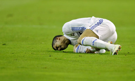
Cristiano Ronaldo was announced today Ballon d'Or and it made me remember something I read some time ago about football players and their injuries. Most of football players injuries are assessed with medical imaging. Medical Imaging provides information about the extent of the injury (e.g. involvement of tendons) and also information about the recovery. The most important exams are: MRI and Ultrasound. Some clubs even have their own ultrasound machine. However, it is still recognized among experts that medical imaging for sports injuries needs further research, because at times it still does not have high sensitivity.

 Cristiano Ronaldo feels the pain as Real Madrid slump to defeat at Osasuna

So how many times did Cristiano Ronaldo undergo an MRI exam?
Ate least three that I could find:
November 2013
August 2010
October 2009

More information here and there:

Woods C, Hawkins RD, Maltby S, Hulse M, Thomas A, Hodson A, & Football Association Medical Research Programme (2004). The Football Association Medical Research Programme: an audit of injuries in professional football--analysis of hamstring injuries. British journal of sports medicine, 38 (1), 36-41 PMID: 14751943

Sunday, July 28, 2013

MRI safety week

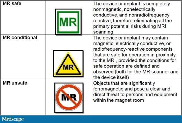
This last week, it was MRI safety week. This event was created to remember the unfortunate death of Michael Columbini, a young boy of 6 years old, caused by a oxygen tank flying in the MRI scanner room. This accident took place in 2001. Even tough everyone working on MRI scanner rooms should know about MRI safety, it is important to renew your knowledge every once in a while. In my institute, MRI safety tests have to be done every 2 years. Major concerns are metal objects, tattoos, wire positioning and SAR. Another very important thing is to keep the scanner rooms clean and organized, something that we researchers tend to forget...

A simple test for you to take is here: http://www.falckproductions.com/mri-safety-week-2013/


I suggest here a review about MRI safety, but I am deeply concerned that there is no more available material online for free. This review has to be payed. Do you know more MRI safety resources?
Mary F Dempsey, Barrie Condon, Donald M Hadley (2002). MRI safety review Seminars in Ultrasound, CT and MRI DOI: 10.1016/S0887-2171(02)90010-7

Wednesday, July 24, 2013

Best/Worse Movie Trailer involving MRI!

This trailer is the most funny/weird/amazing/strange trailer I have ever seen using MRI! The movie looks so bad, that it might be good. I have mixed feelings about seeing this movie... I present you the trailer of "The Drone Virus":


PS - And no, there are no virus in MRI scanners! 

Sunday, June 2, 2013

Procrastination to find the most cited paper in the field of MRI

I have posted recently about the most cited (important?) papers in Medical Imaging in the last ten/five/two years here. Today I look for the most cited papers in the field of MRI. Interesting to note that these 3 papers were published in Neuroimage.

Most cited paper in Radiology, Nuclear Science and Medical Imaging Field about MRI:

- of the last 10 years with 1346 citations:
Ashburner, J., & Friston, K. (2005). Unified segmentation NeuroImage, 26 (3), 839-851 DOI: 10.1016/j.neuroimage.2005.02.018 

This paper is the basis for the SPM framework, one of the most important in the field of MRI. Thus, it is understandable that this paper has a lot of citations, because most researchers who use this framework (and there are a lot, myself included) use this paper in their citations.

of the last 5 years with 250 citations:

Klein, A., Andersson, J., Ardekani, B., Ashburner, J., Avants, B., Chiang, M., Christensen, G., Collins, D., Gee, J., Hellier, P., Song, J., Jenkinson, M., Lepage, C., Rueckert, D., Thompson, P., Vercauteren, T., Woods, R., Mann, J., & Parsey, R. (2009). Evaluation of 14 nonlinear deformation algorithms applied to human brain MRI registration NeuroImage, 46 (3), 786-802 DOI: 10.1016/j.neuroimage.2008.12.037

I have to say that I am quite surprised by finding this paper on top. Brain MRI registration is nowadays considered almost a solved problem and I don't think there are many people looking into this anymore. However, it is always nice to put in your own paper: "I used this registration, because this paper says it is the best".


- of the last 2 years with 106 citations:

Smith, S., Miller, K., Salimi-Khorshidi, G., Webster, M., Beckmann, C., Nichols, T., Ramsey, J., & Woolrich, M. (2011). Network modelling methods for FMRI NeuroImage, 54 (2), 875-891 DOI: 10.1016/j.neuroimage.2010.08.063

I have talked about brain networks some times in this blog and this paper shows me that this topic has been hot in the last two years. This papers discusses different methods to obtain networks with fMRI data: "Many different methods are being used in the literature, but almost none has been carefully validated or compared for use on FMRI timeseries data."

Thursday, May 30, 2013

ESMRMB 2013 - Abstract Submission

This year's deadline for abstract submission is 30th May, so there is no time left to submit. In October, the conference will be in Toulouse, France. Since this is the 30th ESMRMB conference, there will be special surprises according to the organizers. Some preliminary information about the programme is already online.
More information here.


Don't forget that next year, ESMRMB will be held together with ISMRM in Milan and the deadline will be in November. Keep track of your conferences with the Medical Imaging Google Calendar.

Tuesday, April 30, 2013

What does the Muse CD cover have to do with Medical Imaging?

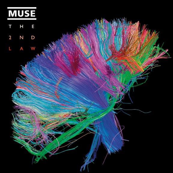
This is Muse CD cover of their 6th album: "The 2nd Law":

Indeed, this image is truly beautiful and remarkable. It is an image of the white matter fibers in the brain obtained with diffusion MRI (link). The image was obtained by the Human Connectome Project, which is a 5-year project funded by NIH to find the networks of the human brain. These networks will show how our brain communicates between different regions and give insight about the anatomical and functional organization of the brain. The project also has the goal to produce data that will help understanding brain diseases such as Alzheimer's disease. The data is available to the scientific community.

So how do you obtain these networks? By applying computer algorithms to data obtained with different neuroimaging techniques: MRI, fMRI, diffusion MRI among others. These computer algorithms come from the graph theory. The application of these algorithms is extremely useful, because the algorithms analyze the network, reduce the complexity, find similarities and differences between different networks.

A very nice science article for researchers not familiar with the topic:
http://www.sciencemag.org/site/products/lst_20130118.xhtml

To know more about obtaining diffusion MRI data or network methods, look into these two articles:
- Hasan, K., Walimuni, I., Abid, H., & Hahn, K. (2011). A review of diffusion tensor magnetic resonance imaging computational methods and software tools Computers in Biology and Medicine, 41 (12), 1062-1072 DOI: 10.1016/j.compbiomed.2010.10.008
- Kaiser, M. (2011). A tutorial in connectome analysis: Topological and spatial features of brain networks NeuroImage, 57 (3), 892-907 DOI: 10.1016/j.neuroimage.2011.05.025

Monday, April 22, 2013

ISMRM 2013

ISMRM 2013 has just started. This annual meeting organized by the International Society for Magnetic Resonance in Medicine is considered by many the most important meeting in the MRI field.


The plenary talks gives us a good idea of the main/hot topics this year:
22.April: Beyond Fourier Encoding: The Need, the Challenges & the Rewards of Breaking Out of K-Space
23.April: MRI in Cancer: Promises, Controversies & Technical Innovation in Breast MRI
24.April: Standardization of MR-Based Biomarkers for Evidence Based Medicine Across Institutions
25.April: MR-PET
26.April: Connectomics: A New Frontier in Neuroscience

What do you think are going to be the hot topics this year?

More information about the program can be found here. Unfortunately, there is no way to follow closely this meeting (there is only twitter #ismrm2013). Can't wait for the time where you can follow conferences online too via video (even if not real-time).


Monday, April 8, 2013

Sculptures inspired by medical imaging

Cao Hiu is a Chinese artist, who took the art of sculptures to the next level. The outside of the sculpture looks like ancient Greek sculptures, but the inside looks just like the inside of a body. He used MRI references to build the interior of the sculpture. Not only these sculptures look interesting and accurate, they can also be quite educational...

Cao Hui (7)

More images can be found here.

Tuesday, April 2, 2013

Best April's Fools Joke in Medical Imaging

This year, Radiopaedia.org, which presents one case per day, presented the best April's fool Joke (1st April) in medical imaging. The case was presented as a real one as it shows here (now it has warnings that it was a joke):
http://radiopaedia.org/cases/april-fools-2013-vertebral-aplasia-von-schlapp-syndrome

The images look pretty crazy, but have amazing realism... (X-ray and MRI respectively):


I have to say that I googled Von Schlapp in the hope of finding about this strange disease, but didn't find anything... but I never taught the case wasn't real. Nevertheless, I am not a doctor... The making of the images can be seen in a video here.

Saturday, March 16, 2013

Procrastination with MRI "Pornography"

This week I have come across the "strangest" MRI videos. The video consists of an MRI acquisition of a couple having sex. The "interesting" part comes after minute 1:30. My first comment was: I can't understand where the actual "interface" between the two people is in some positions. We truly become "One"! Technically I think this must not be easy, because there is not so much space inside the bore and because the couple might not feel very comfortable to do it in such conditions...

Thursday, March 14, 2013

Best MRI books


I have talked before of medical imaging books in this post. Today I make a compilation of the MRI books I have found quite good:

Magnetic Resonance Imaging: Physical Principles and Sequence Design. E. M. Haacke, R. W. Brown, M. R. Thompson, R. Venkatesan.


Handbook of MRI Pulse Sequences. M. Bernstein, K. King, X. J. Zhou.

MRI in Practive. C. Westbrook, C. K. Roth, J. Talbot.

Handbook of MRI Technique. C. Westbrook.

MRI: basic Principles and Applications. M. Brown and R. C. Semelka. 

MRI from Picture to Proton. D. McRobbie, E. Moore, M. Graves, M. Prince.

I think the last four are best to start and the first two are best when your MRI knowledge is already quite good. Do you have other suggestions?

Wednesday, March 13, 2013

Refurbished imaging systems


This company Block Imaging provides refurbished imaging systems. Not always a new system needs to be bought... And you can also find a list of equipments they are looking for here. The video on top is one of their cute/funny promotion videos.

Tuesday, March 12, 2013

Is high-field MRI harmful?

Although high-field MRI has been already tested in humans and considered safe to do clinical studies, there is some new evidence concerning high-field MRI. Apparently, there is a link between depression and the high-field MRI radiographers. I was very surprised by this link. The complete news is here.

My personal thought is that working with high-field MRI might cause depression, not because of the high-field itself, but because working with high-field MRI can be a constant "this is not working that well"! Imagine to work with a machine that is constantly giving problems... I would be depressed too!

I hope there is more clarification about this matter in the future. It is time that we know the long term effects of MRI fields.

Friday, February 22, 2013

Sivu MRI video

A new MRI video has been going around in the internet. Sivu, an english singer, made his latest video clip inside the MRI machine while performing. MRI images were acquired in a hospital in London.  Impressive images were created while he sings:


Sivu - Better Man Than He. from Adam Powell on Vimeo.

The idea for the music video came from CLEFT and Barts and the London Charity, two charities working on treatments for cleft lips and palates in children.

Sunday, February 17, 2013

Great moments in TV with medical images/scanners! #4

ER was another famous medical series. Mark Greene, one of the doctors, realizes that his tumor is back and goes to the MRI to get checked. Notice the detail that he really took all his metallic accessories...

http://www.youtube.com/watch?v=uxhcEY6Y0pk&feature=youtu.be&t=3m20s

P.S. - This video wasn't available for embedding.

Friday, February 15, 2013

Best Medical Imaging Book

A great book is a precious help. I have several medical imaging books, which I use to cross information. General medical imaging books which focus on the physics and engineering principles are my favorites. Here is some of them:

Medical Imaging Physics. William Hendee and E. Russel Ritenour.
http://www.amazon.com/Medical-Imaging-Physics-William-Hendee/dp/0471382264

Medical Imaging Signals and Systems. Jerry Prince and Jonathan Links.
http://www.amazon.com/Medical-Imaging-Signals-Systems-Prince/dp/0130653535

The Essential Physics of Medical Imaging. J. Bushberg, J. Seibert, E. Leidholdt Jr., J. Boone.
http://www.amazon.com/Essential-Physics-Medical-Imaging-Third/dp/0781780578

These two are also quite famous and used, but I have personally never used them:

Introduction to Medical Imaging: Physics, Engineering and Clinical Applications. N. Smith and A. Webb.
http://www.amazon.com/Introduction-Medical-Imaging-Engineering-Applications/dp/0521190657

Fundamentals of Medical Imaging. Paul Suetens.
http://www.amazon.com/Fundamentals-Medical-Imaging-Paul-Suetens/dp/0521519152

Do you know any other good books?

Thursday, February 14, 2013

Valentine's day and medical imaging

Because today is Valentine's day, I show you a video I saw last year which I really liked. Can we really see love in our brains?


The Love Competition from Brent Hoff on Vimeo.