Tuesday, April 30, 2013

What does the Muse CD cover have to do with Medical Imaging?

This is Muse CD cover of their 6th album: "The 2nd Law":

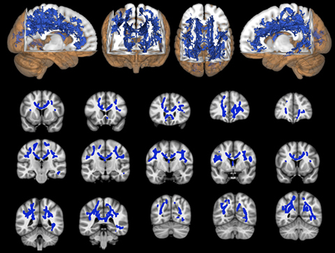
Indeed, this image is truly beautiful and remarkable. It is an image of the white matter fibers in the brain obtained with diffusion MRI (link). The image was obtained by the Human Connectome Project, which is a 5-year project funded by NIH to find the networks of the human brain. These networks will show how our brain communicates between different regions and give insight about the anatomical and functional organization of the brain. The project also has the goal to produce data that will help understanding brain diseases such as Alzheimer's disease. The data is available to the scientific community.

So how do you obtain these networks? By applying computer algorithms to data obtained with different neuroimaging techniques: MRI, fMRI, diffusion MRI among others. These computer algorithms come from the graph theory. The application of these algorithms is extremely useful, because the algorithms analyze the network, reduce the complexity, find similarities and differences between different networks.

A very nice science article for researchers not familiar with the topic:
http://www.sciencemag.org/site/products/lst_20130118.xhtml

To know more about obtaining diffusion MRI data or network methods, look into these two articles:
- Hasan, K., Walimuni, I., Abid, H., & Hahn, K. (2011). A review of diffusion tensor magnetic resonance imaging computational methods and software tools Computers in Biology and Medicine, 41 (12), 1062-1072 DOI: 10.1016/j.compbiomed.2010.10.008
- Kaiser, M. (2011). A tutorial in connectome analysis: Topological and spatial features of brain networks NeuroImage, 57 (3), 892-907 DOI: 10.1016/j.neuroimage.2011.05.025

Monday, April 29, 2013

CT scans help Anthropology studies

This video has caught my attention few days ago. Field Museum in Chicago has used a CT scan to help them with the face reconstruction of an ancient skull (12,000 to 15,000 year old). The great advantage of using a CT scan is that once you have scanned the skull, you can do the processing and reconstruction of the skull on a computer without having to touch it, which means that the skull is never damaged. The problem here is usually the transport of the skull to the CT scan, which are usually located in hospital or clinics. This time, they used a mobile CT scan, which travels to the museum instead of the skull having to travel. Moreover, the researchers used a 3D printer to obtain a model of the skull, which was then used by a forensic artist to do the final face reconstruction. The motivation behind this new scan was that the researchers were not happy with the face reconstruction done before, which made this skull look "too Neanderthal".

After watching this video, I have look for publications which describe the techniques used, but I haven't found many detailed publications. However, I found this review publication (Lynnerup, N. (2010). Medical Imaging of Mummies and Bog Bodies – A Mini-Review Gerontology, 56 (5), 441-448 DOI: 10.1159/000266031), which I have read and although not too detailed about the CT scanning procedures, I have learned a few things:
- Living tissues require different processing that dead tissues, because Hounsfield units are different. Bone tends to de-mineralize, while soft tissues tend to become denser (probably due to surrounding minerals).
- Even that this type of scans are done since the 70's, new CT scans are also done, because technology has improved that new findings arise.
- CT data can be easily shared with other scientists, while skulls and mummies can't.


Saturday, April 27, 2013

Procrastination to find the most cited papers in Medical Imaging

Few day ago, I was wondering what were the most cited (important?) papers in Medical Imaging in the last ten/five/two years. The problem was that I didn't know exactly how to find this information. I googled a bit around and I found a way and tried it out. I found also some extra information about the subject:

Published Papers in Radiology, Nuclear Science and Medical Imaging Field:

Citations in Radiology, Nuclear Science and Medical Imaging Field:

It is interesting to note that it is increasing steadily every year. 

Most cited paper in Radiology, Nuclear Science and Medical Imaging Field:

- of the last 10 years
Jan, S., Santin, G., Strul, D., Staelens, S., Assié, K., Autret, D., Avner, S., Barbier, R., Bardiès, M., Bloomfield, P., Brasse, D., Breton, V., Bruyndonckx, P., Buvat, I., Chatziioannou, A., Choi, Y., Chung, Y., Comtat, C., Donnarieix, D., Ferrer, L., Glick, S., Groiselle, C., Guez, D., Honore, P., Kerhoas-Cavata, S., Kirov, A., Kohli, V., Koole, M., Krieguer, M., Laan, D., Lamare, F., Largeron, G., Lartizien, C., Lazaro, D., Maas, M., Maigne, L., Mayet, F., Melot, F., Merheb, C., Pennacchio, E., Perez, J., Pietrzyk, U., Rannou, F., Rey, M., Schaart, D., Schmidtlein, C., Simon, L., Song, T., Vieira, J., Visvikis, D., Walle, R., Wieërs, E., & Morel, C. (2004). GATE: a simulation toolkit for PET and SPECT Physics in Medicine and Biology, 49 (19), 4543-4561 DOI: 10.1088/0031-9155/49/19/007

- of the last 5 years
Klein, S., Staring, M., Murphy, K., Viergever, M., & Pluim, J. (2010). elastix: A Toolbox for Intensity-Based Medical Image Registration IEEE Transactions on Medical Imaging, 29 (1), 196-205 DOI: 10.1109/TMI.2009.2035616


- of the last 2 years
Hricak, H., Brenner, D., Adelstein, S., Frush, D., Hall, E., Howell, R., McCollough, C., Mettler, F., Pearce, M., Suleiman, O., Thrall, J., & Wagner, L. (2010). Managing Radiation Use in Medical Imaging: A Multifaceted Challenge Radiology, 258 (3), 889-905 DOI: 10.1148/radiol.10101157

How to find this information?
Go to Web of Knowledge and follow these tutorial:
http://www.umuc.edu/library/libhow/woshighlycited_tutorial.cfm

Friday, April 26, 2013

The best blogs in Neuroscience

With the new community of Research Blogging, I have come across with 4 blogs, which are probably the most famous and known in the neuroscience blogosphere of research blogging:


Image from here.

The Scicurious Brain: the Good, Bad, and  Weird in Psychology and Neuroscience and my recommended articles are:
- Beer, Dopamine and Brain Scans
- The BRAIN initiative: BAM or BUST?

Neuroskeptic and my recommended articles are:
- The Man with Uncrossed Eyes
- What is wrong with "Publish or Perish"?

Neurocritic and my recommended articles are:
How Neuroscientists Scan the Media
- What Is This Thing Called Neuroscience?

Brain Posts and my recommendation articles are:
- DTI identifies Brain Aging Changes
- Blast-related Traumatic Brain Injury

These blogs are from this moment in the blog list on the right side of this blog. Did you already know these blogs? Or know other very good ones?

Thursday, April 25, 2013

Research Blogging

This blog is now part of the community in researchblogging.org. "ResearchBlogging.org is a system for identifying the best, most thoughtful blog posts about peer-reviewed research." Know Your Images is now one of the blogs which ResearchBlogging checks for posts about peer-reviewed articles. Furthermore, it is a great way to find new blogs and also keeps you updated about the blogs you like.

Of course, this blog will keep a fare amount of posts which are not dedicated to peer-reviewed articles, but which are just fun to know about and watch.

Wednesday, April 24, 2013

More art inspired by Medical imaging

Marilene Oliver is another artist who is inspired by medical images. She creates sculptures and installations based on medical images. The full portfolio can be seen here. I show in this post some images of her most interesting work:


Tuesday, April 23, 2013

fMRI lie detection and the Semrau case

Semrau is a psychologist accused of committing fraud to Medicare and Medicaid. The case became mostly famous, because he asked that fMRI lie detection would be a evidence in court. The judge had to decide if fMRI was admissible and after hearing scientists advocating for both sides, he has decided not to admit such evidence. However, the question is: Will it be possible to use fMRI lie detection one day?, because the reason for not admitting it has been based on the error rates and acceptance by scientific community and that can change any day...


http://dericbownds.net/uploaded_images/duped.jpg
Image from here

So how does fMRI lie detection work at the moment?
- A deception task is presented to the volunteers: they have to lie about the object they have taken from a box (or similar, such as a card from envelope).
- The volunteer goes inside the scanner and structural MRI is performed and a motor task can also be performed to make the volunteers more familiar with the MRI itself.
- The deception task starts and the volunteer is asked questions about the stolen object among other questions. The volunteer has to lie about stealing the object. During this time, EPI (Echo Planar Imaging) images are acquired.
- Processing of data starts, which includes reorientation and motion correction. Brain patterns are analyzed to detect lying.

Findings have shown that there are specific activated areas (anterior cingulate and the prefrontal cortex) in subjects in the task of deception when a group study is performed. This is a group study, but for fMRI to become a lie detector, it has to stand in individual studies. This has been difficult, because fMRI is a technique with a low signal-to-noise ratio, but some studies have been done. Moreover, deception tasks in these studies are still simple ones, while more complex ones (like the Semrau case) have not been performed.

One of the studies which presented results on individual basis (the one referenced here at the bottom) has led that a company has been formed to sell this type of service (CEPHOS). This was the company involved in the Semrau case and the CEO of this company is the scientist advocating for the fMRI lie detection. The two scientists which advocated against the fMRI lie detector were Marc Raichle, PhD (Wash. U. St. Louis, Neuroscience) and Peter Imrey, PhD (Cleveland Clinic, Statistics).

Anyway, my personal belief is that fMRI should be on the service of health and not of law...

Other Links:
http://www.sciencemag.org/content/328/5984/1336.1.full
http://news.sciencemag.org/scienceinsider/2010/05/fmri-lie-detection-gets-its-day-.html
http://blogs.law.stanford.edu/lawandbiosciences/2012/09/07/hot-news-6th-circuit-affirms-in-us-v-semrau-says-no-to-fmri-lie-detection/

Kozel, F., Johnson, K., Mu, Q., Grenesko, E., Laken, S., & George, M. (2005). Detecting Deception Using Functional Magnetic Resonance Imaging Biological Psychiatry, 58 (8), 605-613 DOI: 10.1016/j.biopsych.2005.07.040

Monday, April 22, 2013

ISMRM 2013

ISMRM 2013 has just started. This annual meeting organized by the International Society for Magnetic Resonance in Medicine is considered by many the most important meeting in the MRI field.


The plenary talks gives us a good idea of the main/hot topics this year:
22.April: Beyond Fourier Encoding: The Need, the Challenges & the Rewards of Breaking Out of K-Space
23.April: MRI in Cancer: Promises, Controversies & Technical Innovation in Breast MRI
24.April: Standardization of MR-Based Biomarkers for Evidence Based Medicine Across Institutions
25.April: MR-PET
26.April: Connectomics: A New Frontier in Neuroscience

What do you think are going to be the hot topics this year?

More information about the program can be found here. Unfortunately, there is no way to follow closely this meeting (there is only twitter #ismrm2013). Can't wait for the time where you can follow conferences online too via video (even if not real-time).


Saturday, April 20, 2013

Procrastination with Coursera

Coursera is a place where you can take online courses from some of the best universities of the world (EPFL, Duke University...) and more universities are joining every month. You just have to register for the course and follow it weekly (on your own schedule). You might have assignments and everything. This is all completely free. There are so many courses that I think I didn't look at them all. Nevertheless, I point you to some that are interesting:

Medical Neuroscience
Healthcare Innovation and Entrepreneurship
Writing in the Sciences
Image and Video Processing
Digital Image Processing
Computational Methods for Data Analysis
High Performance Scientific Computing
Introduction to Data Science

Friday, April 19, 2013

PhD comics talks about Open Access

And because there is no such thing as "too much talk about Open Access":

Original video here.

Thursday, April 18, 2013

The Dead Salmon fMRI experiment

Have you ever heard about the dead salmon fMRI experiment? Me neither, until recently. The study may seem like a waste of money and expertise, but this study has actually open the eyes of the community about one important aspect of fMRI studies: multiple comparisons.

fmri-salmonImage from here.

The basic experiment was just to put a dead salmon inside the MRI scanner, perform an fMRI study and analyze results. Since in an fMRI experiment, you have a lot of voxels being compared, there are some voxels that will eventually show up as activated. In the dead salmon fMRI experiment, voxels in the brain and spine showed up as being activated (see image above). Some could say that even if the salmon is dead, there is activity in its brain, but the most likely (scientific) explanation is that those results just happened by chance. To avoid this problems, you should use a multiple comparison correction, which takes into account this problem. After this correction, of course, the dead salmon showed no activated voxels. Although scientifically based, this study stands out for being so funny. It actually won an IgNobel prize in 2012. The original poster is too good to be true:
methods
Image from here.

Original poster: http://prefrontal.org/files/posters/Bennett-Salmon-2009.pdf
And posts by the author in his blog: http://prefrontal.org/blog/2009/06/atlantic-salmon-index/

Other Links:
http://www.wired.com/wiredscience/2009/09/fmrisalmon/
http://blogs.scientificamerican.com/scicurious-brain/2012/09/25/ignobel-prize-in-neuroscience-the-dead-salmon-study/
http://boingboing.net/2012/10/02/what-a-dead-fish-can-teach-you.html

Wednesday, April 17, 2013

NFL (National Football League) and medical imaging

I have recently talked about the brain injuries in Superbowl players here and there are updated news about this. NFL (National Football League) is supporting a deeper study about these injuries and also how to prevent them. Some are afraid that this will kill the practice of football, but I think it is important that NFL starts taking these health issues seriously. Eventually, this research can even result in broader scientific knowledge.


Image from here.

Links:
http://www.reuters.com/article/2013/03/11/us-nfl-concussion-idUSBRE92A0UT20130311
http://edition.cnn.com/2013/01/29/health/nfl-harvard-study
http://www.newyorker.com/online/blogs/elements/2013/04/how-damaged-are-nfl-players-brains.html


Tuesday, April 16, 2013

TEDMED 2013

I hope you have already heard about TED before (if not, seriously check it out here). TEDMED is a TED conference dedicated only to medical issues and it starts today. I hope medical imaging will be part of some talks...

The schedule of the conference is here, but most important it is possible to watch the conference online in real-time or on-demand following a couple of instructions. I'll see it later on-demand according to my interests and time. Right now, the talks I am looking forward are:
- Can Science take the next leap? by Francis Collins
- What's the 21st century version of prevention? by Danny Hillis
- How can we improve health care if doctors don't know if they do any good? by Michael E. Porter

The TEDMED experience:

The videos from last year can be accessed online in TEDMED youtube channel.

Sunday, April 14, 2013

Great moments in TV with medical images/scanners! #12

This week, I have a great video for you. In the last episode of Modern Family, Claire goes to the hospital to make a check-up cardiac angiography. I was surprised that she stayed in a hospital bed before and after the procedure, so I googled a bit about it. Then, I come across the best angiography video ever! In "The exorcist" (1973), a angiography is performed to Reagen (the main actress) because she is acting "strange". The scene was considered by the director to be the most disturbing one from the whole movie and the medical team there is a real one! However, the procedure is not a real one... (and it is a cerebral angiography).


And the making of:

Friday, April 12, 2013

Making the Brain transparent with Clarity Gel

Recently, there has been news about a new method for neuroimaging. It consists of making the brain "transparent". This new method is probably going to revolutionize imaging of biological tissues. When I first read the news, I wasn't sure how the method really worked. As I have studied in vivo neuroimaging techniques, I also thought that this method was in vivo (they were talking about rats and an autistic brain). So I was wondering how they look through the cranium and also how the biological processes are affected by this new technique. Until I read an article which explained clearly that this is an in vitro technique (Guardian Neurophilosophy Blog by Mo Costandi).


So this new technique is a method to image biological tissues post mortem. The brain passes through a process where it is put into preparation gels and liquids, and after that it is ready to be properly imaged in a electron microscope (or other techniques such as fluorescence microscope), without the need of slicing it. The innovation here is the use of the new Clarity gel, which holds everything together, while fat components are eliminated (fat is what prevents the brain from being imaged correctly). 

Nature video which shows spectacular images using nice visualization techniques:

Other Links:
http://www.nature.com/news/see-through-brains-clarify-connections-1.12768
http://www.abc.net.au/science/articles/2013/04/11/3734738.htm

Thursday, April 11, 2013

Marie Curie Fellowships

If you are about to finish your PhD or have just finished, you can consider applying to a Marie Curie Fellowship. The main idea of these fellowships is to support researchers to move to a European country to perform top research. The next deadlines are in August, but if you are really serious about this, you should start now.

Here is some advice about these fellowships:
1. Read carefully the documentation to find out if you eligible and what you will need. 
2. Start preparing the proposal early enough: contact your host institution and supervisor in charge and then start writing!
3. Don't be afraid to market yourself in the proposal and make sure you have something written about every evaluation point. 
4. Ask many people to review it: from the field and outside the field, the National Contact Points can also review it...
5. Get the support letters to your proposal (3 if possible).

6. If possible, get your hands in somebody else proposal from previous years...


Official Information can be found here, and unofficial information can be found here.
Advices to write a good proposal here.

Wednesday, April 10, 2013

ISBI 2013

ISBI 2013 - International Symposium on Biomedical Imaging - is taking place this week in San Francisco. The complete program is available here and abstracts can be read. The final papers will be available in IEEExplore soon (no date was announced). Unfortunately, this conference has no way to follow it more closely for those who couldn't attend.

Tuesday, April 9, 2013

Scientists read your dreams!?

This week, news have reported that scientists can read your dreams. Examples are here and here, and a more scientific and older one here. First all, just be aware that the titles "Scientists can read dreams" is exaggerated. It is not like they are watching a video of what you are dreaming... What the scientists actually can do is to "read" your brain looking at specific categorized information. Then, scientists "read" your brain while you are sleeping and wake you up to categorize those "readings". They "read" the brains with fMRI and EEG (a well established technology to study sleep disorders). Then, they compared the information and try to guess what the person is dreaming with the information that have collected awake. It is like the awake patterns build a database for the sleeping patterns. The scientists reported “We built a model to predict whether each category of content was present in the dreams”, so not exactly watching a video of your dreams... Now, when they put together this experiment with these one, we will have videos of our dreams!

Image copied from here.

Second thing I would like to point out is that while it is interesting for research to study dreams and sleep, I am not sure this research is really good news for everybody... I personally don't want anyone looking into my dreams... I hope this topic stays inside the boundaries of research.

Monday, April 8, 2013

Sculptures inspired by medical imaging

Cao Hiu is a Chinese artist, who took the art of sculptures to the next level. The outside of the sculpture looks like ancient Greek sculptures, but the inside looks just like the inside of a body. He used MRI references to build the interior of the sculpture. Not only these sculptures look interesting and accurate, they can also be quite educational...

Cao Hui (7)

More images can be found here.

Sunday, April 7, 2013

Great moments in TV with medical images/scanners! #11

Nurse Jackie tries to trick her best friend O'Hara to give her medication and gets caught... and she uses medical images in her deception.

Saturday, April 6, 2013

Procrastination with Scientific Social Networks


I have written before about Google Scholar as a way to follow your co-authors, colleagues or just to get alerts from a certain topic. Today, I talk about other websites that have nice features which Google Scholar doesn't have and which can be useful: BiomedExperts, ResearchGate and Academia.edu.


BiomedExperts is a scientific social network for biomedical experts.The main features of this webpage are the visualization tools about researchers. You can see the published topics, published articles, co-authors, times and places, network view and geonetwork view. These two last ones are very nice to visualize and give you information that Google Scholar doesn't provide you.


ResearchGate is another scientific network, but it is available for all research topics. It has basically all the features of BiomedExperts, expect the network visualization, but it has other ones, such as citation management, endorsement management (like LinkedIn) and even allows you to publish data (or download it).


Academia.edu is another scientific network with a lot of users. The main difference from the other two is that it allows you to put papers which are not finished or were not accepted. The thing I most like in this webpage is that it gives me an alert whenever my profile appears in google results, so basically I know when other researchers googled me and which words they used for that.

This post was an idea from a fellow researcher.