Wednesday, February 27, 2013

Siemens Molecular Imaging

Do you want to know more about Siemens Molecular Imaging?

Check the products here:
http://healthcare.siemens.com/molecular-imaging
and the customer portal here:
http://healthcare.siemens.com/molecular-imaging/customer-portal-resource/administration

I found the growth resources in this page quite interesting, specially the white papers.

Sunday, February 24, 2013

Saturday, February 23, 2013

Procrastination with IEEE

IEEE is the Institute of Electrical and Electronics Engineers (IEEE, read I-Triple-E). The good thing about procrastination with IEEE is that you don't feel that bad, because it is work related. IEEE spectrum is the magazine for the members and there is also a version online: http://spectrum.ieee.org/biomedical/imaging
I found this article very interesting in IEEE spectrum and I recommend:
http://spectrum.ieee.org/tech-talk/biomedical/imaging/medical-imaging-group-calls-for-fewer-ct-tests-and-more-research-on-health-effects

And also there are seminars available online:
http://spectrum.ieee.org/webinar/

Friday, February 22, 2013

Sivu MRI video

A new MRI video has been going around in the internet. Sivu, an english singer, made his latest video clip inside the MRI machine while performing. MRI images were acquired in a hospital in London.  Impressive images were created while he sings:


Sivu - Better Man Than He. from Adam Powell on Vimeo.

The idea for the music video came from CLEFT and Barts and the London Charity, two charities working on treatments for cleft lips and palates in children.

Thursday, February 21, 2013

Stanford Mini Med School

I have recently come across these videos from Stanford medical school, where an introduction about several topics of health and research are talked about. The list of videos can be found here: http://www.youtube.com/course?list=ECD7B0DBD8C28BDEA2 

I post here the one about medical imaging:


Wednesday, February 20, 2013

Gorillla CT scan

Can you find the Gorilla in this image?

Notice anything off?

I took at least 10 seconds to spot it. This image was used in a study with radiologists, where they were asked if there is something wrong with the image. 83% of radiologist didn't see the gorilla. This doesn't mean that the radiologist don't pay attention to the images, but rather that they will not see what they aren't looking for... Please see the video that inspired this study in the links and understand better what this means.

Links:
http://www.theinvisiblegorilla.com/videos.html
http://life.nationalpost.com/2013/02/13/dancing-gorilla-image-on-ct-scan-passed-over-by-more-than-80-of-radiologists-study-finds/


Tuesday, February 19, 2013

Open-Source frameworks for medical imaging

C++ frameworks are great to develop new methods from other previously coded methods. Here are some great frameworks for medical imaging:

ITK Insight Segmentation and Registration Toolkit http://www.itk.org/
As the name states, it is great for segmentation and registration, but also has filters, statistics...

STIR Software for Tomographic Image Reconstruction http://stir.sourceforge.net/
It is a open-source framework to reconstruct data for nuclear medicine.

ROOT Rapid Object Oriented  Data Framework http://root.cern.ch/
It is one of the major open source codes ever developed for Physics, so it is great to use in medical physics oriented medical imaging.

XIP eXtensible Imaging Platform   http://www.openxip.org/
https://wiki.nci.nih.gov/display/XIP/eXtensible+Imaging+Platform+-+XIP
A platform design for rapid development of medical applications including DICOM tools, renders...

MITK Medical Imaging Toolkit http://www.mitk.org/
A framework which comprises ITK and VTK, a visualization toolkit.

A complete list with more frameworks:
http://www0.cs.ucl.ac.uk/opensource_mia_ws_2012/links.html

Monday, February 18, 2013

Databases of Medical Images

Medical Image Databases are great to work on medical imaging processing and sometimes to get a feeling of the images before using real data. I have found some databases in the internet that I consider quite interesting:


Minimal Interval Resonance Imaging in Alzheimer's Disease MIRIAD
This database is focused on Alzheimer Diseases patients, but also control patients.

Mini Mammographic Image Analysis Society Database Mini-MIAS
A database with mammograms and with medical information.

Medpix Teaching database MedPix
A database with examples to teach from CT to MRI.

Retrospective Image registration Evaluation RIRE
A database with images to register and the ground truth to compare.

Alzheimer's Disease Neuroimaging Initiative ADNI
This a huge database of patients images with PET and MRI.

Do you know other good databases?


Sunday, February 17, 2013

Great moments in TV with medical images/scanners! #4

ER was another famous medical series. Mark Greene, one of the doctors, realizes that his tumor is back and goes to the MRI to get checked. Notice the detail that he really took all his metallic accessories...

http://www.youtube.com/watch?v=uxhcEY6Y0pk&feature=youtu.be&t=3m20s

P.S. - This video wasn't available for embedding.

Saturday, February 16, 2013

Procrastination with Google Scholar

When you don't feel like doing any real work, you can procrastinate with Google Scholar Profiles. Either to find some papers from your favorite authors or just to update your profile.

Why and How to get a Google profile: http://scholar.google.com/intl/en/scholar/citations.html

Here is some examples of Google scholar profiles with lots of citations and papers:
http://scholar.google.com/citations?user=9fGrB0sAAAAJ&hl=en
http://scholar.google.com/citations?user=jHEgTSwAAAAJ&hl=en
http://scholar.google.com/citations?user=CeajmrUAAAAJ&hl=en

And some Medical Imaging Journals:
http://scholar.google.com/citations?hl=en&view_op=search_venues&vq=medical+imaging


Friday, February 15, 2013

Best Medical Imaging Book

A great book is a precious help. I have several medical imaging books, which I use to cross information. General medical imaging books which focus on the physics and engineering principles are my favorites. Here is some of them:

Medical Imaging Physics. William Hendee and E. Russel Ritenour.
http://www.amazon.com/Medical-Imaging-Physics-William-Hendee/dp/0471382264

Medical Imaging Signals and Systems. Jerry Prince and Jonathan Links.
http://www.amazon.com/Medical-Imaging-Signals-Systems-Prince/dp/0130653535

The Essential Physics of Medical Imaging. J. Bushberg, J. Seibert, E. Leidholdt Jr., J. Boone.
http://www.amazon.com/Essential-Physics-Medical-Imaging-Third/dp/0781780578

These two are also quite famous and used, but I have personally never used them:

Introduction to Medical Imaging: Physics, Engineering and Clinical Applications. N. Smith and A. Webb.
http://www.amazon.com/Introduction-Medical-Imaging-Engineering-Applications/dp/0521190657

Fundamentals of Medical Imaging. Paul Suetens.
http://www.amazon.com/Fundamentals-Medical-Imaging-Paul-Suetens/dp/0521519152

Do you know any other good books?

Thursday, February 14, 2013

Valentine's day and medical imaging

Because today is Valentine's day, I show you a video I saw last year which I really liked. Can we really see love in our brains?


The Love Competition from Brent Hoff on Vimeo.

Wednesday, February 13, 2013

Open Source Medical Record

When we get sick, we want to get the best medical treatment possible, specially when our case is a difficult one. This is what Salvatore Iaconesi did. He release his medical files in the internet in the hope to get more and better advice about his brain tumor. Furthermore  he cracked the medical files and converted to open formats so that everyone could use them. He had surgery this month and he is at the moment fine.

Is this the beginning of something new in medical imaging?


Links:
http://artisopensource.net/cure/
http://blog.ted.com/2012/10/02/how-salvatore-iaconesi-has-started-a-movement-for-open-source-medical-files/

Tuesday, February 12, 2013

Time-of-Flight PET vs. MRA

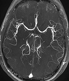
Every time I hear Time-of-Flight TOF, I get confused because it can actually mean TOF PET or TOF MRA. The two acronyms have the some words: Time Of Flight, but have different meanings. 

TOF PET - This PET technology means that the time difference between two photons detected as a coincidence will be used to estimate the actual place of positron annihilation.

TOF MRA - This Magnetic Resonance angiography technique means that a sequence with small time of repetition TR will be used to saturate the stationary nucleus, while the flowing nucleus with Time-Of-Flight will be maximal. 

Both techniques produce beautiful images:


More information on TOF-PET:
http://miil.stanford.edu/research/tofdetector.html

More information on TOF-MRA: 
http://www.imaios.com/en/e-Courses/e-MRI/MR-Angiography-Flow-imaging/time-of-flight-mra 

Monday, February 11, 2013

Simulation for Nuclear Medicine

Simulation is a very important tool in medical imaging, as I already wrote in a previous post. In nuclear medicine, Monte-Carlo simulation is the most used method to simulate data. There  are  several  Monte-Carlo packages, which vary in precision and accuracy of  the simulated effects and computational demand.  Some research groups devised their own Monte-Carlo computer codes, built on top of a general-purpose code, which serves as core layer and application-specific modules are constructed in a hierarchical layer architecture.

PET-EGS based on EGS4 (Electron Gamma Shower 4) http://irs.inms.nrc.ca/research/imaging/
GATE (Geant4 Application for Tomographic Emission) http://www.opengatecollaboration.org/

Dedicated packages that have supported PET and/or SPECT research are:
SimSET (Simulation System for Emission Tomography)http://depts.washington.edu/simset/html/user_guide/user_guide_index.html
SORTEO (Simulation Of Realistic Tridimensional Emitting Objects) http://sorteo.cermep.fr/
PeneloPET (PENetration and Energy LOss of PET) http://nuclear.fis.ucm.es/penelopet/

GATE is one of the most famous packages and the paper published by the authors is here:
http://iopscience.iop.org/0031-9155/49/19/007/

Do you recommend other packages?

Friday, February 8, 2013

Radiation dose in medical imaging

During this week, the French doctors in the case of radiation overdose were convicted and I was surprised to read that between 2001 and 2006, at least 400 patients underwent about 8% more radiation than the safer dose. This was mainly because radiation dose in radiological exams prior to treatment was not taken into account. I always thought that medical professionals were aware that several medical imaging exams expose the patient to radiation dose. An extra mechanism for signaling was developed in France after this case.

Aunt Minnie article:
http://www.auntminnieeurope.com/index.aspx?sec=nws&sub=rad&pag=dis&ItemID=607680&wf=1
Guardian U.K. article:
http://www.guardian.co.uk/world/2012/sep/24/french-doctors-trial-cancer-overdose

A quick internet tool gives you radiation dose of typical exams: http://www.xrayrisk.com/calculator/calculator-normal-studies.php
For imaging professionals, there is more information on radiation dose:
http://www.imagewisely.org/

Thursday, February 7, 2013

MRI courses 2013

This week, I have received the new list of ESMRMB courses for 2013. This courses are a great way to upgrade your skills and knowledge. I, myself, am thinking of taking the Advanced Neuro Imaging: Diffusion, Perfusion, Spectroscopy.

Links for School of MRI: http://www.esmrmb.org/index.php?id=/en/school_of_mri.htm
Links for Lectures of MRI: http://www.esmrmb.org/index.php?id=/en/lectures_on_mr/courses_2013
Links for Hands-On MRI: http://www.esmrmb.org/index.php?id=/en/index/hands-on_mri.htm

ISMRM courses: http://www.ismrm.org/meetings-workshops/future-ismrm-workshops/

Other interesting courses:
Erasmus Course on MRI: http://www.emricourse.org/index.html

Basic Knowledge courses:
MRI education (with Catherine Westbrook): http://www.mrieducation.com/courses/index.html

Wednesday, February 6, 2013

Superbowl and medical imaging?

And because the superbowl was this weekend, I draw your attention to a recent study made with former NFL players. PET studies with FDDNP tracer, developed for Alzheimer disease (AD), have been performed and showed differences between normal subjects and the former NFL players. Nevertheless, the study group is small and therefore, more subjects are necessary to make a bolder statement.

PET plus FDDNP shows brain injury in retired NFL players


Full article about the study:
FDDNP-PET Shows Brain Injury in Former NFL Players
The Journal of Nuclear Medicine article about FDDNP and PIB tracers for AD  
http://jnm.snmjournals.org/content/50/2/191.short

Tuesday, February 5, 2013

The Human Brain Project

The European Commission has officially announced the selection of the Human Brain Project (HBP) as one of its two FET (Future and Emerging Technologies) Flagship projects. The new project will coordinate European efforts to address one of the greatest challenges of modern science: understanding the human brain.

"Understanding the human brain is one of the greatest challenges facing 21st century science. If we can rise to the challenge, we can gain profound insights into what makes us human, develop new treatments for brain diseases and build revolutionary new computing technologies."


"According to a recent report, nearly one-third of the European population is affected by some kind of psychiatric or neurological disease (anxiety, mood disorders, neurodegenerative disease, etc.)."

The promoting video is very instructive and I'm very exciting about the database with medical images, of course... I hope this project will be very successful!


Monday, February 4, 2013

Breast tomosynthesis

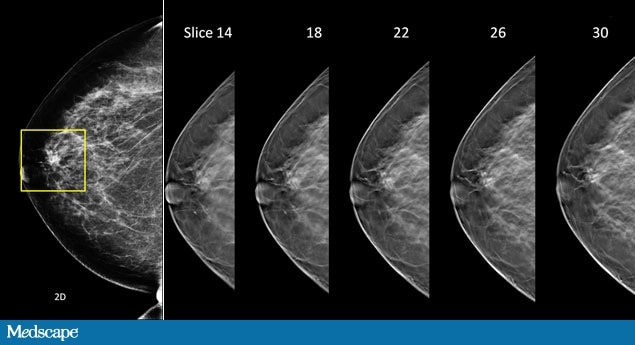
Breast tomosynthesis is a 3D technique, which I believe will change the screening of breast cancer. This technique is able to produce 3D images of the breast, instead of 2D projection images available in mammography. 3D imaging can unfold small lesions, such as micro-calcification and give a better understanding of the 3D shape of the lesion. Furthermore, it is possible that in the future compression will be less than in the mammography and dose will be comparable.


Tomosynthesis for the general public:
http://www.massgeneral.org/imaging/services/3D_mammography_tomosynthesis.aspx
Tomosynthesis for researchers: http://online.medphys.org/a_review_of_breast_tomosynthesis_part_i_the_image_acquisition_process?mode=www
http://online.medphys.org/resource/1/mphya6/v40/i1/p014302_s1

About breast compression:
http://www.ncbi.nlm.nih.gov/pubmed/20228049

Saturday, February 2, 2013

Procrastination #3

And because today is Saturday, I give you some more procrastination sources:

A Programmer's life (a cartoon based on real events!): http://aprogrammerslife.info/
Khan academy brain teasers: https://www.khanacademy.org/math/brain-teasers


Friday, February 1, 2013

Simulation MRI - free tools

Simulation is essential when working on medical imaging from the technique development point of view. Therefore, tools to do MRI simulation are described here. All of these tools have scientific papers published which you can cite in your work.

Brainweb http://brainweb.bic.mni.mcgill.ca/brainweb/
This tools works mostly as a database of brain MRI's with specific sequences and artifacts, but several parameters can be manipulated to obtain custom made simulations.

JEMRIS http://www.jemris.org/
This tool allows to define the phantom and the sequence, therefore it is one of the most flexible tools available. The reconstruction of non-linear sequences is not available.

MRI Simulation and Reconstruction http://bigwww.epfl.ch/algorithms/mri-reconstruction/
This package was developed in Matlab and allows to simulate and reconstruct datasets.

SIMRI http://www.simri.org/
This package is able to simulate several advanced artifacts and sequences, although the package itself does not seem very intuitive.

FSL POSSUM http://fsl.fmrib.ox.ac.uk/fsl/fsl-4.1.9/possum/index.html
This is a part of the well-know MRI/fMRI software FSL, which allows simulation of MRI images with some artifacts.

Do you know other MRI simulators? Please share.