Sunday, July 28, 2013

MRI safety week

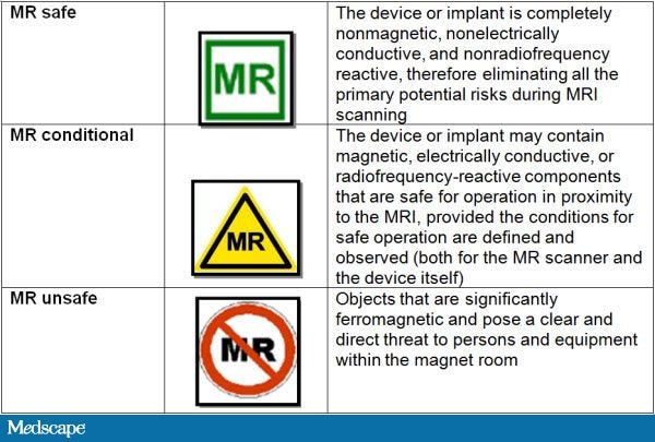
This last week, it was MRI safety week. This event was created to remember the unfortunate death of Michael Columbini, a young boy of 6 years old, caused by a oxygen tank flying in the MRI scanner room. This accident took place in 2001. Even tough everyone working on MRI scanner rooms should know about MRI safety, it is important to renew your knowledge every once in a while. In my institute, MRI safety tests have to be done every 2 years. Major concerns are metal objects, tattoos, wire positioning and SAR. Another very important thing is to keep the scanner rooms clean and organized, something that we researchers tend to forget...

A simple test for you to take is here: http://www.falckproductions.com/mri-safety-week-2013/


I suggest here a review about MRI safety, but I am deeply concerned that there is no more available material online for free. This review has to be payed. Do you know more MRI safety resources?
Mary F Dempsey, Barrie Condon, Donald M Hadley (2002). MRI safety review Seminars in Ultrasound, CT and MRI DOI: 10.1016/S0887-2171(02)90010-7

Wednesday, July 24, 2013

Best/Worse Movie Trailer involving MRI!

This trailer is the most funny/weird/amazing/strange trailer I have ever seen using MRI! The movie looks so bad, that it might be good. I have mixed feelings about seeing this movie... I present you the trailer of "The Drone Virus":


PS - And no, there are no virus in MRI scanners! 

Monday, July 22, 2013

ERC Starting Grant Winners

ERC awards the most important grants to researchers in Europe. For young researchers (2-7 years after PhD), the ERC has Starting Grants. The goal for these starting grants is for these young researchers to start their own group and line of research. This year results have been published (here) and of course, I checked which of these grants are related to medical imaging. There were 3329 applications and 287 researchers were successful. The ERC grants are divided in three big areas: 1) Physical Sciences and Engineering, 2) Life Sciences, 3) Social Sciences and Humanities. Medical imaging fits in Life Sciences and more concretely in LS7 - Diagnostic tools. There were 8 starting grants related to Medical Imaging:

Daniela Thorwarth, Erlangen University, Germany - Biologically individualized, model-based  radiotherapy on the basis of multi-parametric molecular tumour profiling.

Julien Valette, CEA, France - Exploring brain intracellular space using diffusion-weighted NMR spectroscopy in vivo

Galia Blum, The Hebrew University of Jerusalem, Israel - Protease Activated X-Ray Contrast Agents for 
Molecular Imaging of Vulnerable Atherosclerotic Plaques and Cancer Development using Spectral CT.

Rachel Katz-Brull, Hadassah Medical Organization, Israel - Citicoline and deoxyglucose as new molecular imaging probes of DNP hyperpolarized MRI for cancer and neuroimaging

Niv Papo, Ben-Gurion University of the Negev, Israel - Developing Multispecific Biological Agents that
Target Tumor Neovasculature for Cancer Imaging and Therapy

Francesco Ricci, University of Rome "Tor Vergata", Italy - Nature-inspired theranostic nanodevices for tumor imaging, early diagnosis and targeted drug-release

Mangala Srinivas, Radboud University Nijmegen, The Netherlands - Clinical ultrasound platform for the quantitative and longitudinal imaging of theranostics and cellular therapy

Jaco Zwanenburg, University Medical Centre, Utrecht, The Netherlands - Towards understanding cerebral small vessel disease: Innovative, MRI-based, functional markers to discover the terra incognita between large vessels and macroscopic brain lesions

Congratulations to all the winners! But I still have a favorite grant and therefore, one of the papers related to that grant can be read here:

Daniela Thorwarth, Susanne-Martina Eschmann, Frank Paulsen, Markus Alber. (2006). Hypoxia Dose Painting by Numbers: A Planning Study International Journal of Radiation Oncology * Biology * Physics DOI: 10.1016/j.ijrobp.2006.11.061


Thursday, July 18, 2013

Right Dose Image Contest (by Siemens)

Every year, Siemens organizes a Medical Imaging Contest. The idea is to obtain the best CT image with minimal dose. This contest doesn't focus only on image quality, but also on the dose, which is displayed in top of every image. There are always very nice images from different body parts and the competition this year is already open. Just take a look at the gallery. Of course, as it is organized by Siemens, the images have to be acquired with a scanner from the SOMATOM® Family and post-processed with syngo® software. Learn more about the challenge:

http://healthcare.siemens.com/computed-tomography/technologies-innovations/ct-image-contest


To get more information about radiation in CT, read this article:
Mannudeep K. Kalra, MD, DNB, Michael M. Maher, MD, FFR(RCSI), FRCR, Thomas L. Toth, DSc, Leena M. Hamberg, PhD, Michael A. Blake, MRCPI, FRCR, FFR(RCSI), Jo-Anne Shepard, MD and Sanjay Saini, MD (2004). Strategies for CT Radiation Dose Optimization Radiology DOI: 10.1148/radiol.2303021726

Tuesday, July 16, 2013

Statistics - Love or Hate?

There is no clinical medical imaging paper without statistics. Research can't live without it, but most researchers wish they could. Big Picture is a Welcome Trust magazine, which is directed to teachers and students. Therefore, the magazine has really simple and clear examples. The new issue is about statistics. There is lots of information relevant for medical imaging researchers: 1) the story about the advertisement of a CT scan that was forbidden to broadcast, 2) types of medical studies from the statistic point of view (see picture below) and 3) general concepts about research. See more information here.


The new issue pdf can be read here. The previous issue was about medical neuroimaging, which can be read here.


Monday, July 15, 2013

Best posts in the first 6 months - from Angelina Jolie to Muse

This blog has just turned 6 months. I make the balance of these first six months and compare it with the first three months:

Top 5 posts (4 new entries, but #1 post is still the same):
1. Medical Imaging Conferences 2013
2. How does a painting look like on X-ray?
3. What does the Muse CD cover have to do with Medical Imaging?
5. Dissecting Art, Intersecting Anatomy - Medical illustration

More than 9400 visualizations (6900 in the last three months, which is more than the double of the first three months) and 136 posts (53 new posts, 60% of the first three months)! Thank you for sharing this blog!


Friday, July 12, 2013

Breast Imaging and Computer Aided Diagnosis

When I started studying biomedical engineering and I learned a lit bit about programming, my ingenuity made me think that one day we could build programs that don't need medical doctors. WRONG! Nevertheless, we can still to provide better algorithms and results to medical doctors which make the hard clinical decisions. One of the most challenging tasks for a doctor is to analyze mammograms to find microcalcifications and evaluate the malignity of a lesion. It takes years of experience to be a very good analyst of these images. This is where we can hope to help the medical doctors.

Computer Aided Diagnosis (CADx) and Computer Aided Detection (CAD) are important tools for the medical doctors and a lot of these tools are being investigated and evaluated in the context of breast imaging using mammograms. At the moment, there is still no method which proved to be accurate enough to be brought to clinical practice. More about this can be read in this review:

Ganesan K, Acharya UR, Chua CK, Min LC, Abraham KT, & Ng KH (2013). Computer-aided breast cancer detection using mammograms: a review. IEEE reviews in biomedical engineering, 6, 77-98 PMID: 23247864

Wednesday, July 10, 2013

PET answers: "Does Cannabis make you lazy?"

PET (Positron Emission Tomography) is the image modality in which I have put most of my sweat and tears, so I am always interested to know the latest findings using this modality. The most recent question PET tries to answer is: "Does Cannabis make you lazy?"

To study this, the researchers used "radio-labelled dopamine" ([18F]-DOPA) in a group of cannabis smokers and a group of controls. Dopamine is the chemical in the brain that controls pleasure and motivation (read here). The study found that cannabis smokers (3xweek) had decreased levels of dopamine. Since these levels are decreased, researchers think this will contribute to the lack of "pleasure" of the cannabis smoker and therefore, the smoker will be make less effort to obtain something. And that is why cannabis can make you lazy! This is probably what inspired this song:


The study can be found here.
Bloomfield MA, Morgan CJ, Egerton A, Kapur S, Curran HV, & Howes OD (2013). Dopaminergic Function in Cannabis Users and Its Relationship to Cannabis-Induced Psychotic Symptoms. Biological psychiatry PMID: 23820822

Monday, July 8, 2013

South Korea, here I go!

My latest abstract to a conference was accepted today and I will be attending the conference in South Korea. It will be my first time in Asia... and in Gangnam (style)! More details about the conference "IEEE Nuclear Science Symposium and Medical Imaging Conference" can be seen here: NSS/MIC, but the scientific programme is still not available. Nevertheless, the webpage has already information about the plenary talks, short courses and workshops. Hope to see some of you there!